You have no items in your shopping cart.
Description
Research Area
Signal Transduction
Images & Validation
−Item 1 of 7
| Tested Applications | FC, IF, IHC-P, WB |
|---|---|
| Dilution Range | IF - 1:10-50, WB - 1:16000, IHC-P - 1:10-50, FC - 1:10-50 |
| Reactivity | Human |
Key Properties
−| Host | Rabbit |
|---|---|
| Clonality | Polyclonal |
| Isotype | Rabbit IgG |
| Immunogen | This FASN antibody is generated from rabbits immunized with a KLH conjugated synthetic peptide between 942-973 amino acids from the Central region of human FASN. Antigen Region: 942-973 aa. |
| Target | FASN |
| Molecular Weight | 273427 Da |
| Conjugation | Unconjugated |
Storage & Handling
−| Storage | Maintain refrigerated at 2-8°C for up to 2 weeks. For long term storage store at -20°C in small aliquots to prevent freeze-thaw cycles |
|---|---|
| Form/Appearance | Purified polyclonal antibody supplied in PBS with 0.09% (W/V) sodium azide. This antibody is prepared by Saturated Ammonium Sulfate (SAS) precipitation followed by dialysis against PBS. |
| Expiration Date | 12 months from date of receipt. |
| Disclaimer | For research use only |
Alternative Names
−Fatty acid synthase, [Acyl-carrier-protein] S-acetyltransferase, [Acyl-carrier-protein] S-malonyltransferase, 3-oxoacyl-[acyl-carrier-protein] synthase, 3-oxoacyl-[acyl-carrier-protein] reductase, 3-hydroxyacyl-[acyl-carrier-protein] dehydratase, Enoyl-[acyl-carrier-protein] reductase, Oleoyl-[acyl-carrier-protein] hydrolase, FASN, FAS
Similar Products
−- Item 1 of 7
- Item 1 of 2
FASN Mouse Monoclonal Antibody [orb1473885]
IF, WB
Human
Mouse
Monoclonal
Unconjugated
200 μl, 100 μl, 50 μl, 30 μl - Item 1 of 2
FASN Rabbit Polyclonal Antibody [orb213921]
IF, WB
Human
Rabbit
Polyclonal
Unconjugated
30 μl, 100 μl, 200 μl, 50 μl

Quality Guarantee
Explore bioreagents carefree to elevate your research. All our products are rigorously tested for performance. If a product does not perform as described on its datasheet, our scientific support team will provide expert troubleshooting, a prompt replacement, or a refund. For full details, please see our Terms & Conditions and Buying Guide. Contact us at [email protected].
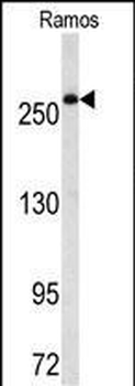
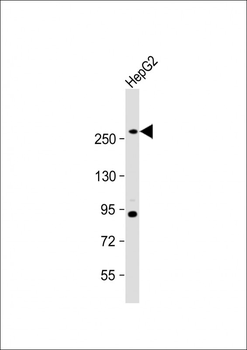
Western blot analysis of FASN Antibody (Center) in Ramos cell line lysates (35 ug/lane).FASN (arrow) was detected using the purified Pab.
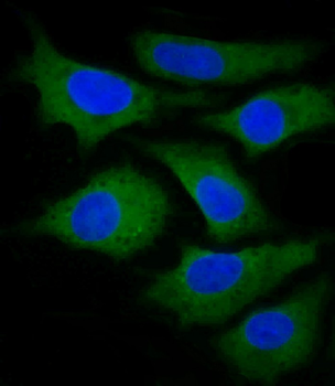
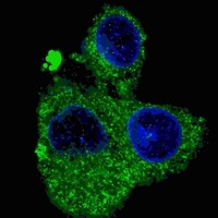
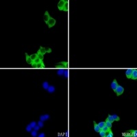
Confocal immunofluorescent analysis of FASN Antibody (Center) with Hela cell followed by Alexa Fluor 488-conjugated goat anti-rabbit lgG (green).DAPI was used to stain the cell nuclear (blue).
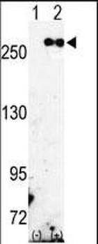
Western blot analysis of FASN (arrow) using rabbit polyclonal FASN Antibody (Center). 293 cell lysates (2 ug/lane) either nontransfected (Lane 1) or transiently transfected with the FASN gene (Lane 2).
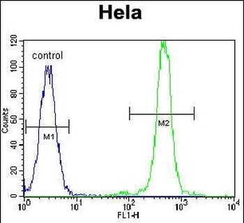
FASN Antibody (Center) flow cytometric analysis of Hela cells (right histogram) compared to a negative control cell (left histogram). FITC-conjugated goat-anti-rabbit secondary antibodies were used for the analysis.
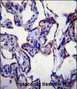
FASN Antibody (Center) immunohistochemistry analysis in formalin fixed and paraffin embedded human placenta tissue followed by peroxidase conjugation of the secondary antibody and DAB staining.This data demonstrates the use of FASN Antibody (Center) for immunohistochemistry. Clinical relevance has not been evaluated.
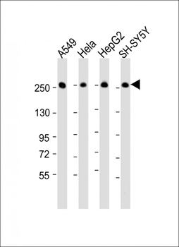
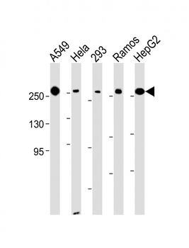
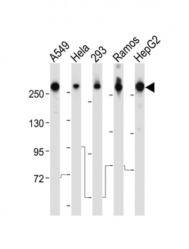
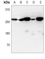
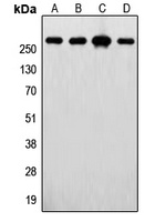
All lanes: Anti-FASN Antibody (Center) at 1: 16000 dilution. Lane 1: A549 whole cell lysate. Lane 2: Hela whole cell lysate. Lane 3: HepG2 whole cell lysate. Lane 4: SH-SY5Y whole cell lysate. Lysates/proteins at 20 µg per lane. Secondary Goat Anti-Rabbit IgG, (H+L), Peroxidase conjugated at 1/10000 dilution. Predicted band size: 273 kDa. Blocking/Dilution buffer: 5% NFDM/TBST.
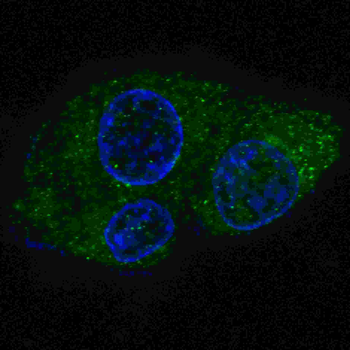
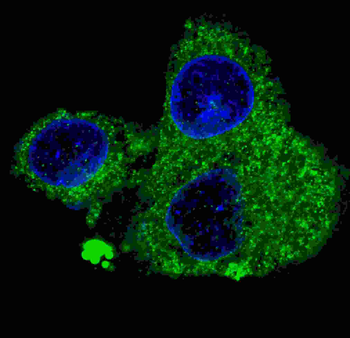
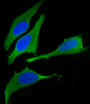
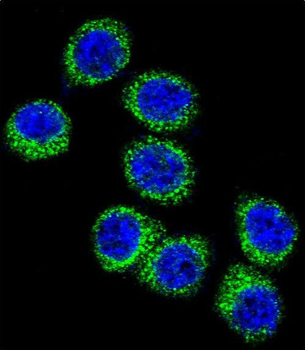
Fluorescent confocal image of HepG2 cells stained with FASN (Center) antibody. HepG2 cells were fixed with 4% PFA (20 min), permeabilized with Triton X-100 (0.2%, 30 min). Cells were then incubated with FASN (Center) primary antibody (1: 200, 2 h at room temperature). For secondary antibody, Alexa Fluor 488 conjugated donkey anti-rabbit antibody (green) was used (1:1000, 1h). Nuclei were counterstained with Hoechst 33342 (blue) (10 μg/ml, 5 min).
Quick Database Links
UniProt Details
− No UniProt data available
NCBI Reference Sequences
−Associated Accession Numbers
Curated reference sequences for the gene transcript and protein product| Protein | NP_004095.4 |
|---|
Documents Download
Datasheet
Product Information
Request a Document
Protocol Information
WB
Western Blot (IB, immunoblot)
IHC-P
Immunohistochemistry Paraffin
FC
Flow Cytometry
IF
Immunofluorescence
FASN Antibody (Center) (orb1929252)

- 0.0
Based on 0 reviews
Participating in our Biorbyt product reviews program enables you to support fellow scientists by sharing your firsthand experience with our products.
Login to Submit a ReviewAvailable Sizes
Select a size below
Free Secondary Antibody (20 ul)0/0
Please add an antibody product to your cart first.