You have no items in your shopping cart.
Description
Research Area
Epigenetics & Chromatin
Images & Validation
−Item 1 of 7
| Tested Applications | FC, IF, IHC-P, WB |
|---|---|
| Dilution Range | IF - 1:25, WB - 1:2000, IHC-P - 1:10-50, FC - 1:25 |
| Reactivity | Human, Mouse |
Key Properties
−| Host | Rabbit |
|---|---|
| Clonality | Polyclonal |
| Isotype | Rabbit IgG |
| Immunogen | This EZH2 antibody is generated from rabbits immunized with a recombinant fragment (N-term) protein from human EZH2. Antigen Region: 1-296 aa. |
| Target | EZH2 (HGNC:3527) |
| Molecular Weight | 85363 Da |
| Conjugation | Unconjugated |
Storage & Handling
−| Storage | Maintain refrigerated at 2-8°C for up to 2 weeks. For long term storage store at -20°C in small aliquots to prevent freeze-thaw cycles |
|---|---|
| Form/Appearance | Purified polyclonal antibody supplied in PBS with 0.09% (W/V) sodium azide. This antibody is purified through a protein A column, followed by peptide affinity purification. |
| Expiration Date | 12 months from date of receipt. |
| Disclaimer | For research use only |
Alternative Names
−Histone-lysine N-methyltransferase EZH2, ENX-1, Enhancer of zeste homolog 2, Lysine N-methyltransferase 6, EZH2, KMT6
Similar Products
−- Item 1 of 7
- Item 1 of 8
- Item 1 of 7
EZH2 Recombinant Rabbit Monoclonal Antibody [orb1151968]
IF, IHC-Fr, IHC-P, WB
Mouse, Rat
Human, Mouse, Rat
Rabbit
Recombinant
Unconjugated
50 μl, 100 μl, 25 μl - Item 1 of 6
- Item 1 of 1
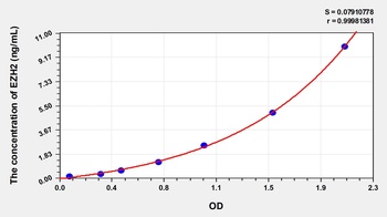
Human Enhancer of Zeste Homolog 2 (EZH2) ELISA Kit [orb777392]
Human
0.16-10 ng/mL
0.061 ng/mL
48 T, 96 T

Quality Guarantee
Explore bioreagents carefree to elevate your research. All our products are rigorously tested for performance. If a product does not perform as described on its datasheet, our scientific support team will provide expert troubleshooting, a prompt replacement, or a refund. For full details, please see our Terms & Conditions and Buying Guide. Contact us at [email protected].
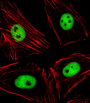
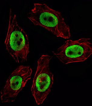
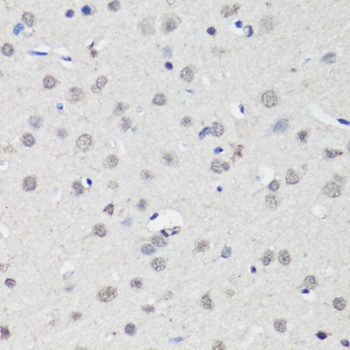
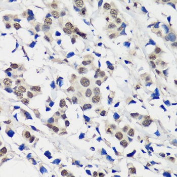
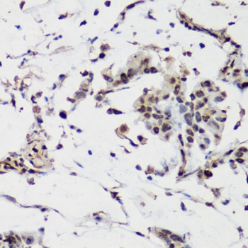
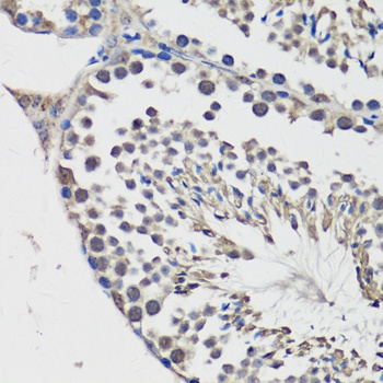
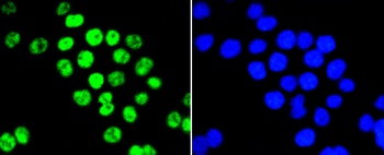
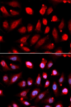
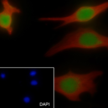
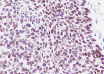
Immunofluorescent analysis of 4% paraformaldehyde-fixed, 0.1% Triton X-100 permeabilized Hela (Human cervical epithelial adenocarcinoma cell line) cells labeling EZH2 at 1/25 dilution, followed by Alexa Fluor 488-conjugated goat anti-rabbit IgG secondary antibody at 1/400 dilution (green). Confocal image showing nuclear staining on Hela cell line. Cytoplasmic actin is detected with Alexa Fluor 555 conjugated with Phalloidin at 1/100 dilution (red).
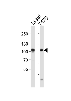
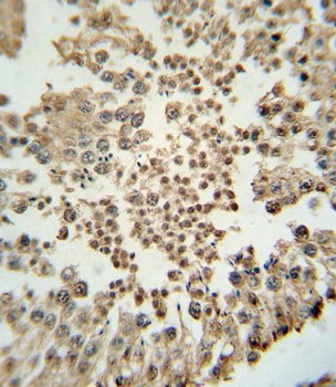
Western blot analysis of lysates from Jurkat, T47D cell line (from left to right), using EZH2 Antibody. Diluted at 1:2000 at each lane. A goat anti-rabbit IgG H&L (HRP) at 1:10000 dilution was used as the secondary antibody. Lysates at 20 ug per lane.

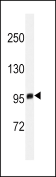
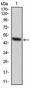
Anti-EZH2 Antibodyat 1:500 dilution + MDA-MB-468 whole cell lysates. Lysates/proteins at 20 µg per lane. Secondary Goat Anti-Rabbit IgG, (H+L), Peroxidase conjugated at 1/10000 dilution. Predicted band size: 85 kDa. Blocking/Dilution buffer: 5% NFDM/TBST.
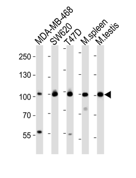
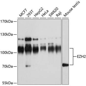
Western blot analysis of lysates from MDA-MB-468, SW620, T47D cell line, mouse spleen, mouse testis tissue (from left to right), using EZH2 Antibody. Diluted at 1:1000 at each lane. A goat anti-rabbit IgG H&L (HRP) at 1:10000 dilution was used as the secondary antibody. Lysates at 20 ug per lane.
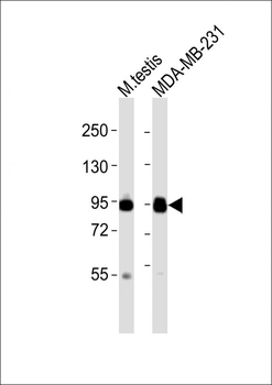
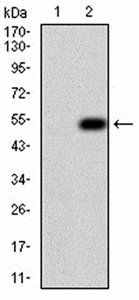
All lanes: Anti-EZH2 Antibody at 1:2000 dilution. Lane 1: mouse testis lysates. Lane 2: MDA-MB-231 whole cell lysates. Lysates/proteins at 20 µg per lane. Secondary Goat Anti-Rabbit IgG, (H+L), Peroxidase conjugated at 1/10000 dilution. Predicted band size: 85 kDa. Blocking/Dilution buffer: 5% NFDM/TBST.
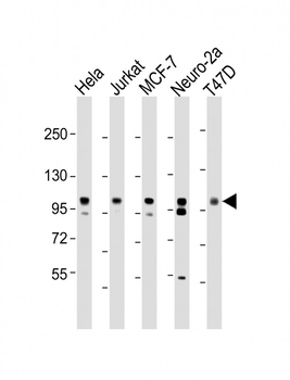
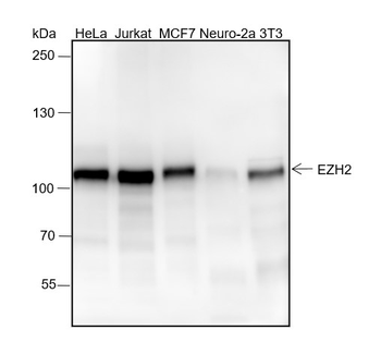
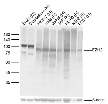
All lanes: Anti-EZH2 Antibody at 1:2000 dilution. Lane 1: Hela whole cell lysate. Lane 2: Jurkat whole cell lysate. Lane 3: MCF-7 whole cell lysate. Lane 4: Neuro-2a whole cell lysate. Lane 5: T47D whole cell lysate. Lysates/proteins at 20 µg per lane. Secondary Goat Anti-Rabbit IgG, (H+L), Peroxidase conjugated at 1/10000 dilution. Predicted band size: 85 kDa. Blocking/Dilution buffer: 5% NFDM/TBST.
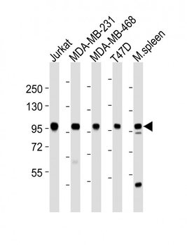
All lanes: Anti-EZH2 Antibody at 1:2000 dilution. Lane 1: Jurkat whole cell lysates. Lane 2: MDA-MB-231 whole cell lysates. Lane 3: MDA-MB-468 whole cell lysates. Lane 4: T47D whole cell lysates. Lane 5: mouse spleen lysates. Lysates/proteins at 20 µg per lane. Secondary Goat Anti-Rabbit IgG, (H+L), Peroxidase conjugated at 1/10000 dilution. Predicted band size: 85 kDa. Blocking/Dilution buffer: 5% NFDM/TBST.
Quick Database Links
Gene Symbol
EZH2 (HGNC:3527)
UniProt
RefSeq (Protein):NP_001190178.1, NP_001190176.1, NP_001190177.1, NP_694543.1, NP_004447.2
UniProt Details
− No UniProt data available
NCBI Reference Sequences
−Associated Accession Numbers
Curated reference sequences for the gene transcript and protein productDocuments Download
Datasheet
Product Information
Request a Document
Protocol Information
WB
Western Blot (IB, immunoblot)
IHC-P
Immunohistochemistry Paraffin
FC
Flow Cytometry
IF
Immunofluorescence
EZH2 Antibody (orb1931591)

- 0.0
Based on 0 reviews
Participating in our Biorbyt product reviews program enables you to support fellow scientists by sharing your firsthand experience with our products.
Login to Submit a ReviewAvailable Sizes
Select a size below
Choose Conjugation or Carrier Free Version
Free Secondary Antibody (20 ul)0/0
Please add an antibody product to your cart first.