You have no items in your shopping cart.
Description
Research Area
Signal Transduction
Images & Validation
−Item 1 of 6
| Tested Applications | FC, IF, IHC-P, WB |
|---|---|
| Dilution Range | IF - 1:10-50, WB - 1:1000, IHC-P-Leica - 1:500, FC - 1:10-50 |
| Reactivity | Human |
Key Properties
−| Antibody Type | Primary Antibody |
|---|---|
| Host | Rabbit |
| Clonality | Polyclonal |
| Isotype | Rabbit IgG |
| Immunogen | This ErbB2 antibody is generated from rabbits immunized with human recombinant ErbB2 protein. |
| Target | ERBB2 |
| Molecular Weight | 137910 Da |
| Conjugation | Unconjugated |
Storage & Handling
−| Storage | Maintain refrigerated at 2-8°C for up to 2 weeks. For long term storage store at -20°C in small aliquots to prevent freeze-thaw cycles |
|---|---|
| Form/Appearance | Purified polyclonal antibody supplied in PBS with 0.09% (W/V) sodium azide. This antibody is purified through a protein A column, followed by peptide affinity purification. |
| Expiration Date | 12 months from date of receipt. |
| Disclaimer | For research use only |
Alternative Names
−Receptor tyrosine-protein kinase erbB-2, Metastatic lymph node gene 19 protein, MLN 19, Proto-oncogene Neu, Proto-oncogene c-ErbB-2, Tyrosine kinase-type cell surface receptor HER2, p185erbB2, CD340, ERBB2, HER2, MLN19, NEU, NGL
Similar Products
−- Item 1 of 4
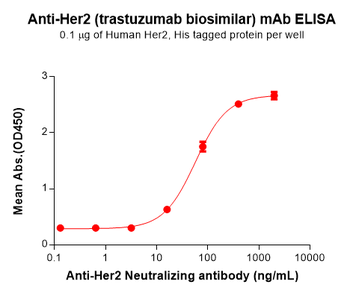
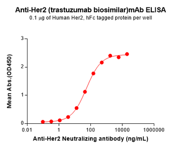
Trastuzumab Biosimilar Antibody, Research Grade (Anti-Her2 / ErbB2) [orb1173652]
ELISA, FC
Human
Human Humanized
Monoclonal
Unconjugated
50 μg, 100 μg - Item 1 of 4
ERBB2 Antibody(N-term) [orb38262]
FC, IHC-P, WB
Mouse, Rat
Human
Rabbit
Polyclonal
Unconjugated
100 μl, 50 μl - Item 1 of 5
C-erbB-2/HER2 Mouse Monoclonal Antibody [orb499961]
IF, IHC-Fr, IHC-P, WB
Mouse, Rat
Human
Mouse
Monoclonal
Unconjugated
50 μl, 100 μl, 200 μl, 200 μg - Item 1 of 4
Neu rabbit pAb Antibody [orb768024]
ELISA, IF, IHC, WB
Human, Mouse, Rat
Polyclonal
Unconjugated
100 μl - Item 1 of 4
Neu rabbit pAb Antibody [orb765798]
ELISA, IF, IHC, WB
Human, Mouse, Rat
Polyclonal
Unconjugated
50 μl, 100 μl

Quality Guarantee
Explore bioreagents carefree to elevate your research. All our products are rigorously tested for performance. If a product does not perform as described on its datasheet, our scientific support team will provide expert troubleshooting, a prompt replacement, or a refund. For full details, please see our Terms & Conditions and Buying Guide. Contact us at [email protected].
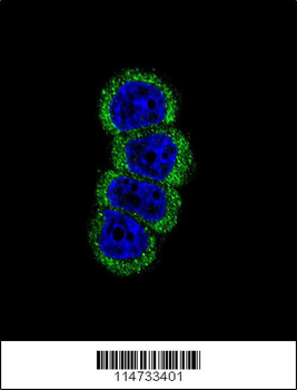
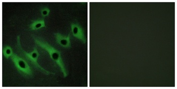
Confocal immunofluorescent analysis of ErbB2 antibody with MCF-7 cell followed by Alexa Fluor 488-conjugated goat anti-rabbit lgG (green). DAPI was used to stain the cell nuclear (blue).
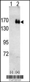
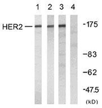
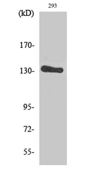
Western blot analysis of HER2 (arrow) using rabbit polyclonal HER2 antibody. 293 cell lysates (2 ug/lane) either nontransfected (Lane 1) or transiently transfected with the HER2 gene (Lane 2).
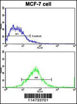
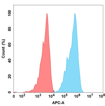
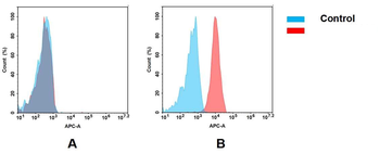
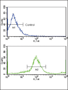
Flow cytometric analysis of MCF-7 cells using HER2 Antibody (bottom histogram) compared to a negative control cell (top histogram). FITC-conjugated goat-anti-rabbit secondary antibodies were used for the analysis.
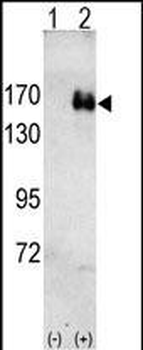
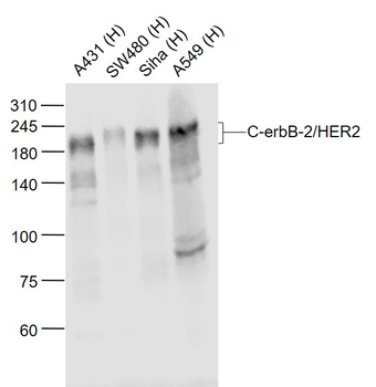
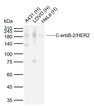
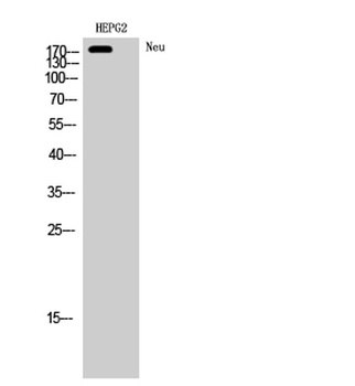
Anti-HER2 Antibody at 1:1000 dilution + SK-BR-3 whole cell lysate. Lysates/proteins at 20 µg per lane. Secondary Goat Anti-Rabbit IgG, (H+L), Peroxidase conjugated at 1/10000 dilution. Predicted band size: 138 kDa. Blocking/Dilution buffer: 5% NFDM/TBST.
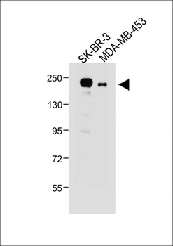
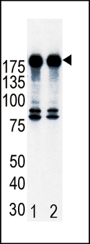
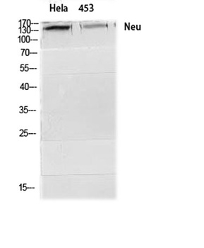
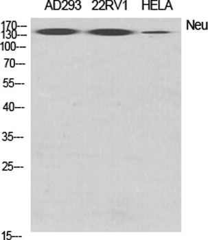
All lanes: Anti-HER2/ERBB2 Antibody at 1:1000 dilution. Lane 1: SK-BR-3 whole cell lysate. Lane 2: MDA-MB-453 whole cell lysate. Lysates/proteins at 20 µg per lane. Secondary Goat Anti-Rabbit IgG, (H+L), Peroxidase conjugated at 1/10000 dilution. Predicted band size: 138 kDa. Blocking/Dilution buffer: 5% NFDM/TBST.
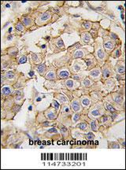
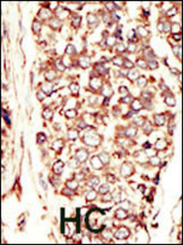
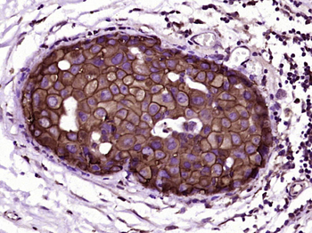
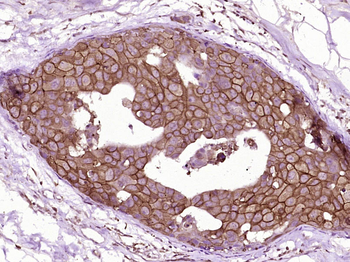
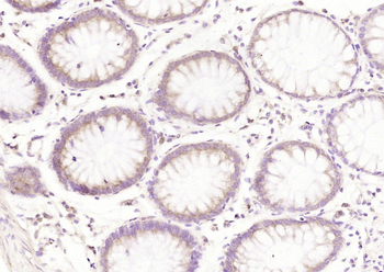
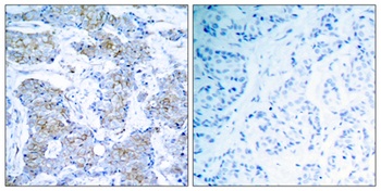
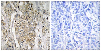
Immunohistochemical analysis of paraffin-embedded Human breast carcinoma tissue was performed on the Leica BOND RXm. Tissue was fixed with formaldehyde at room temperature, antigen retrieval was by heat mediation with a EDTA buffer (pH9.0). Samples were incubated with primary antibody (1:500) for 1 hours at room temperature. A undiluted biotinylated CRF Anti-Polyvalent HRP Polymer antibody was used as the secondary antibody.
Quick Database Links
UniProt Details
− No UniProt data available
NCBI Reference Sequences
−Associated Accession Numbers
Curated reference sequences for the gene transcript and protein product| Protein | NP_001005862.1, NP_004439.2 |
|---|
Documents Download
Datasheet
Product Information
Request a Document
Protocol Information
WB
Western Blot (IB, immunoblot)
IHC-P
Immunohistochemistry Paraffin
FC
Flow Cytometry
IF
Immunofluorescence
ERBB2 Antibody (orb1929061)

- 0.0
Based on 0 reviews
Participating in our Biorbyt product reviews program enables you to support fellow scientists by sharing your firsthand experience with our products.
Login to Submit a ReviewAvailable Sizes
Select a size below
Choose Conjugation or Carrier Free Version
Free Secondary Antibody (20 ul)0/0
Please add an antibody product to your cart first.