You have no items in your shopping cart.
Featured
Description
Research Area
Neuroscience
Images & Validation
−Item 1 of 6
| Tested Applications | IHC-P, WB |
|---|---|
| Reactivity | Human, Mouse |
| Application Notes |
Key Properties
−| Antibody Type | Primary Antibody |
|---|---|
| Host | Rabbit |
| Clonality | Polyclonal |
| Isotype | Rabbit Ig |
| Immunogen | This EphA4 antibody is generated from rabbits immunized with a KLH conjugated synthetic peptide between 875-904 amino acids from the C-terminal region of human EphA4. |
| Target | EPHA4 |
| Molecular Weight | 110 kDa |
| Purification | This antibody is prepared by Saturated Ammonium Sulfate (SAS) precipitation followed by dialysis |
| Conjugation | Unconjugated |
Storage & Handling
−| Storage | Maintain refrigerated at 2-8°C for up to 2 weeks. For long term storage store at -20°C in small aliquots to prevent freeze-thaw cycles. |
|---|---|
| Form/Appearance | Liquid |
| Buffer/Preservatives | Supplied in PBS with 0.09% (W/V) sodium azide. |
| Concentration | batch dependent |
| Expiration Date | 12 months from date of receipt. |
| Disclaimer | For research use only |
Alternative Names
−Ephrin type-A receptor 4, EPH-like kinase 8, EK8, hEK8, Tyrosine-protein kinase TYRO1, Tyrosine-protein kinase receptor SEK, EPHA4, HEK8, SEK, TYRO1
Similar Products
−- Item 1 of 6
EPHA4 Rabbit Polyclonal Antibody [orb507567]
IHC-P, WB
Guinea pig, Human, Mouse, Rat
Rabbit
Polyclonal
Unconjugated
100 μg - Item 1 of 5
EphA4 Antibody (N-term) [orb1929100]
FC, IF, IHC-P, WB
Mouse, Other
Human
Rabbit
Polyclonal
Unconjugated
50 μl, 100 μl - Item 1 of 1
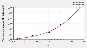
Human Ephrin Type A Receptor 4 (EPHA4) ELISA Kit [orb778905]
Human
0.32-20 ng/mL
0.112 ng/mL
48 T, 96 T - Item 1 of 3
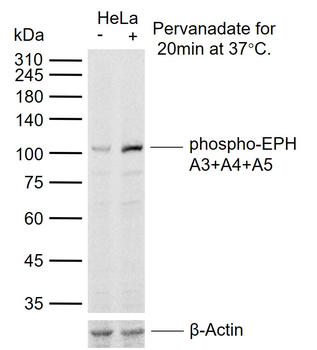
Phospho-EPH A3+A4+A5 (Tyr779) Rabbit Polyclonal Antibody [orb100971]
IF, IHC-Fr, IHC-P, WB
Bovine, Canine, Equine, Gallus, Porcine, Rabbit
Human, Mouse, Rat
Rabbit
Polyclonal
Unconjugated
50 μl, 200 μl, 100 μl - Item 1 of 3

Quality Guarantee
Explore bioreagents carefree to elevate your research. All our products are rigorously tested for performance. If a product does not perform as described on its datasheet, our scientific support team will provide expert troubleshooting, a prompt replacement, or a refund. For full details, please see our Terms & Conditions and Buying Guide. Contact us at [email protected].

Western blot analysis of lysates from Hela, HUVEC cell line (from left to right), using EPHA4 Antibody at 1:1000 at each lane.
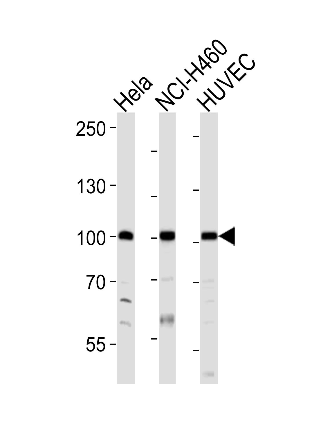
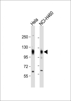
Western blot analysis of lysates from Hela, NCI-H460, HUVEC cell line (from left to right), using EPHA4 Antibody at 1:1000 at each lane.

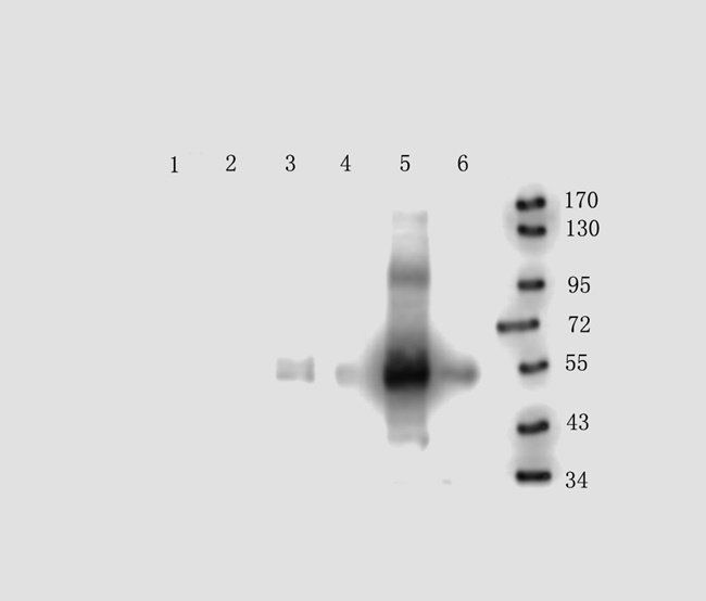
Western blot analysis of lysates from Hela, NCI-H460, mouse NIH/3T3 cell line and human ovary tissue lysate (from left to right), using EPHA4 Antibody at 1:1000 at each lane.
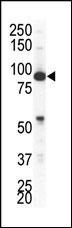
Western blot analysis of anti-EphA4 Pab in NCI-H460 cell lysate
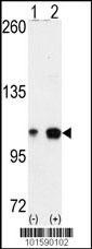
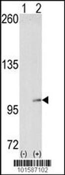
Western blot analysis of EPHA4 using EphA4 Antibody using 293 cell lysates (2 ug/lane) either nontransfected (Lane 1) or transiently transfected with the EPHA4 gene (Lane 2).
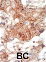
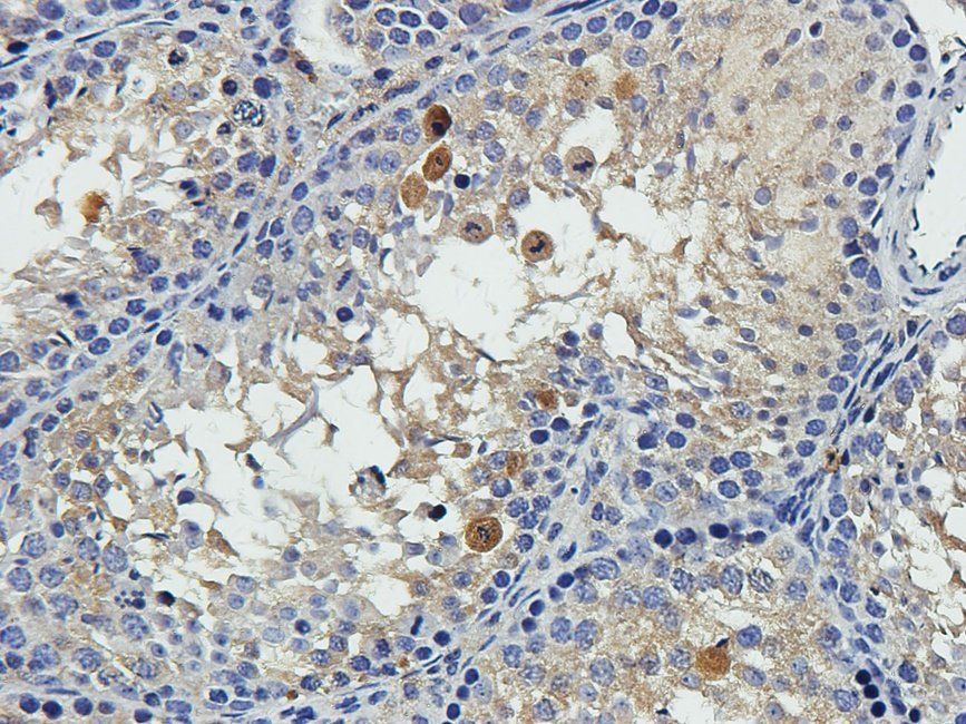
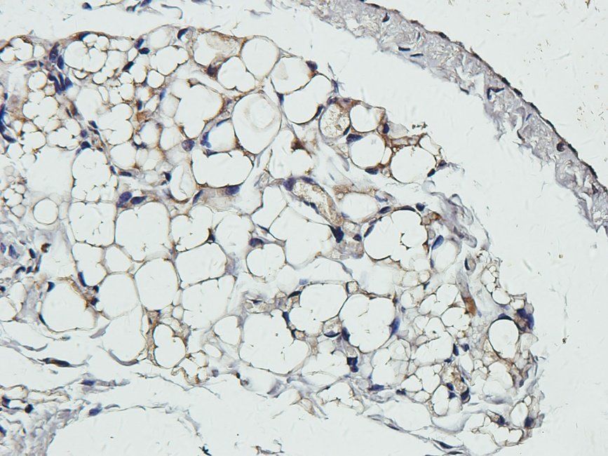
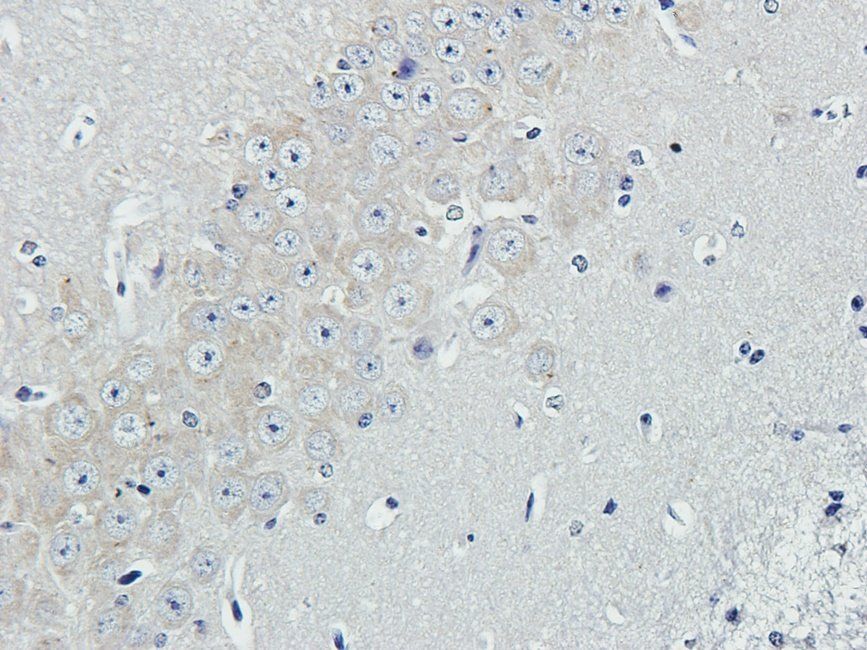
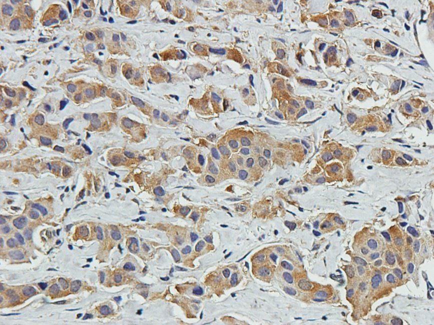
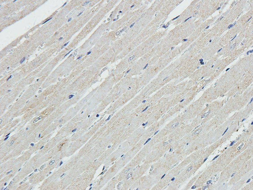
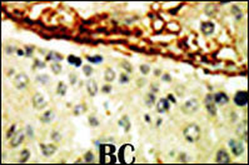
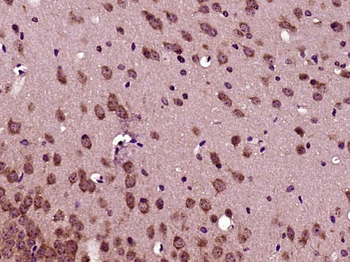
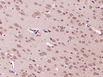
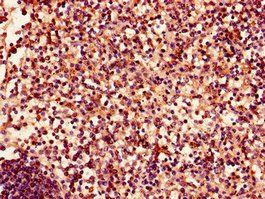
Formalin-fixed and paraffin-embedded human cancer tissue reacted with the primary antibody, which was peroxidase-conjugated to the secondary antibody, followed by DAB staining. BC = breast carcinoma; HC = hepatocarcinoma.
Documents Download
Datasheet
Product Information
Request a Document
Protocol Information
WB
Western Blot (IB, immunoblot)
IHC-P
Immunohistochemistry Paraffin
EPHA4 Antibody (orb1271019)

- 0.0
Based on 0 reviews
Participating in our Biorbyt product reviews program enables you to support fellow scientists by sharing your firsthand experience with our products.
Login to Submit a ReviewAvailable Sizes
Select a size below
Free Secondary Antibody (20 ul)0/0
Please add an antibody product to your cart first.