You have no items in your shopping cart.
Description
Images & Validation
−Item 1 of 5
| Tested Applications | IHC-P, WB |
|---|---|
| Dilution Range | WB: 1:1000, IHC-P: 1:25, IHC-P: 1:25, IHC-P: 1:25, IHC-P: 1:25 |
| Reactivity | Human |
Key Properties
−| Antibody Type | Primary Antibody |
|---|---|
| Host | Mouse |
| Clonality | Monoclonal |
| Isotype | IgG1 |
| Immunogen | This EGFR antibody is generated from mice immunized with a KLH conjugated synthetic peptide between 1163-1191 amino acids from the C-terminal region of human EGFR. Antigen Region: 1163-1191 aa. |
| Target | EGFR (HGNC:3236) |
| Molecular Weight | 134277 Da |
| Conjugation | Unconjugated |
Storage & Handling
−| Storage | Maintain refrigerated at 2-8°C for up to 2 weeks. For long term storage store at -20°C in small aliquots to prevent freeze-thaw cycles |
|---|---|
| Form/Appearance | Purified monoclonal antibody supplied in PBS with 0.09% (W/V) sodium azide. This antibody is purified through a protein G column, followed by dialysis against PBS. |
| Expiration Date | 12 months from date of receipt. |
| Disclaimer | For research use only |
Alternative Names
−Epidermal growth factor receptor, Proto-oncogene c-ErbB-1, Receptor tyrosine-protein kinase erbB-1, EGFR, ERBB, ERBB1, HER1
Similar Products
−- Item 1 of 6
- Item 1 of 3
EGFR-S1026 Antibody (C-term) [orb1930503]
FC, IHC-P, WB
Human
Rabbit
Polyclonal
Unconjugated
50 μl, 100 μl - Item 1 of 3
EGFR Rabbit Polyclonal Antibody [orb338826]
IF, IHC, WB
Human, Mouse
Rabbit
Polyclonal
Unconjugated
30 μl, 100 μl, 200 μl, 50 μl - Item 1 of 3
EGFR Rabbit Polyclonal Antibody [orb304744]
IF, IHC, WB
Human, Monkey, Mouse, Rat
Rabbit
Polyclonal
Unconjugated
30 μl, 100 μl, 200 μl, 50 μl - Item 1 of 2
EGFR Rabbit Polyclonal Antibody [orb213879]
IHC, WB
Human, Monkey, Mouse, Rat
Rabbit
Polyclonal
Unconjugated
30 μl, 100 μl, 200 μl, 50 μl


Quality Guarantee
Explore bioreagents carefree to elevate your research. All our products are rigorously tested for performance. If a product does not perform as described on its datasheet, our scientific support team will provide expert troubleshooting, a prompt replacement, or a refund. For full details, please see our Terms & Conditions and Buying Guide. Contact us at [email protected].
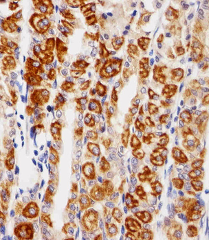
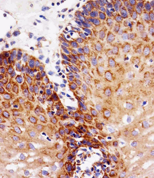
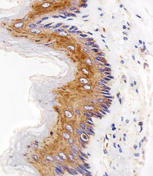
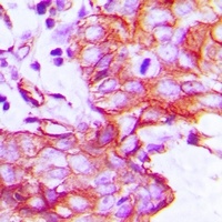
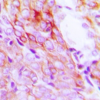
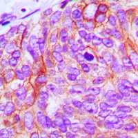
Immunohistochemical analysis of paraffin-embedded H. stomach section using EGFR Antibody (C-term). Diluted at 1:25 dilution. A peroxidase-conjugated goat anti-mouse IgG at 1:400 dilution was used as the secondary antibody, followed by DAB staining.

Immunohistochemical analysis of paraffin-embedded R. esophagus section using EGFR Antibody (C-term). Diluted at 1:25 dilution. A peroxidase-conjugated goat anti-mouse IgG at 1:400 dilution was used as the secondary antibody, followed by DAB staining.
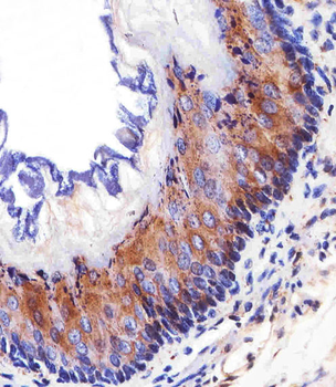
Immunohistochemical analysis of paraffin-embedded M. esophagus section using EGFR Antibody (C-term). Diluted at 1:25 dilution. A peroxidase-conjugated goat anti-mouse IgG at 1:400 dilution was used as the secondary antibody, followed by DAB staining.

Immunohistochemical analysis of paraffin-embedded H. esophagus section using EGFR Antibody (C-term). Diluted at 1:25 dilution. A peroxidase-conjugated goat anti-mouse IgG at 1:400 dilution was used as the secondary antibody, followed by DAB staining.
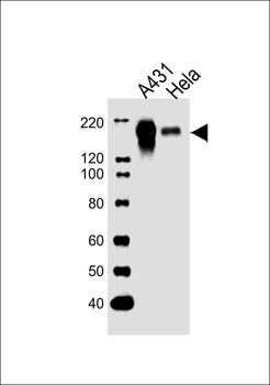
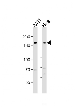
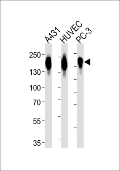
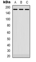
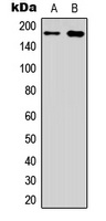
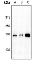
All lanes: Anti-EGFR Antibody (C-term) at 1:1000 dilution. Lane 1: A431 whole cell lysates. Lane 2: Hela whole cell lysates. Lysates/proteins at 20 µg per lane. Secondary Goat Anti-Mouse IgG, (H+L), Peroxidase conjugated at 1/10000 dilution. Predicted band size: 134 kDa. Blocking/Dilution buffer: 5% NFDM/TBST.
Quick Database Links
Gene Symbol
EGFR (HGNC:3236)
UniProt
RefSeq (Protein):NP_958441.1, NP_958439.1, NP_958440.1, NP_005219.2
UniProt Details
− No UniProt data available
NCBI Reference Sequences
−Associated Accession Numbers
Curated reference sequences for the gene transcript and protein product| Protein | NP_958441.1, NP_958439.1, NP_958440.1, NP_005219.2 |
|---|
Documents Download
Datasheet
Product Information
Request a Document
Protocol Information
WB
Western Blot (IB, immunoblot)
IHC-P
Immunohistochemistry Paraffin
EGFR Antibody (C-term) (orb1788111)

- 0.0
Based on 0 reviews
Participating in our Biorbyt product reviews program enables you to support fellow scientists by sharing your firsthand experience with our products.
Login to Submit a Review