
You have no items in your shopping cart.
Description
Research Area
Cancer Biology, Cell Biology, Epigenetics & Chromatin, Molecular Biology, Protein Biochemistry, Signal Transduction
Images & Validation
−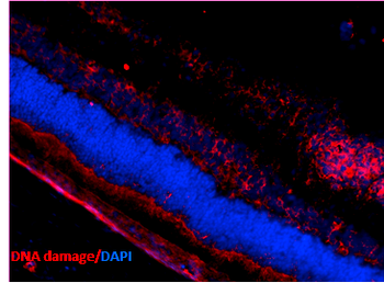
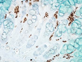
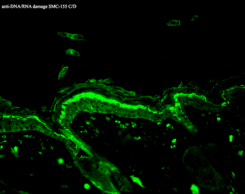
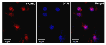
Item 1 of 4
| Tested Applications | DOT, ELISA, FA, FC, ICC, IF, IHC, IP |
|---|---|
| Dilution Range | IHC (1:1000) |
| Reactivity | All |
Key Properties
−| Host | Mouse |
|---|---|
| Clonality | Monoclonal |
| Isotype | IgG2b |
| Clone No. | 15A3 |
| Immunogen | 8-hydroxy-guanosine-BSA and –casein conjugates |
| Target | DNA Damage |
| Purification | Protein G Purified |
| Conjugation | APC |
Storage & Handling
−| Storage | Conjugated antibodies should be stored according to the product label |
|---|---|
| Buffer/Preservatives | 95.46mM Phosphate, 2.48mM MES and 2mM EDTA |
| Concentration | 1 mg/ml |
| Expiration Date | 12 months from date of receipt. |
| Disclaimer | For research use only |
Alternative Names
−8-Hydroxy Guanine, 8-OH-dG, 8OHG, 80G, 8-hydroxyguanine, 8-hydroxy-2'-deoxyguanosine, 8-hydroxyguanosine, 8 OHG, 8-OHG, 8OHdG
Similar Products
−- Item 1 of 3
MSH5 Rabbit Polyclonal Antibody (APC) [orb1002526]
IF
Bovine, Canine, Equine, Human, Porcine, Rabbit, Sheep
Mouse, Rat
Rabbit
Polyclonal
APC
100 μlGTPBP9 Rabbit Polyclonal Antibody (APC) [orb992185]
FC
Bovine, Canine, Drosophila, Rat, Sheep
Human, Mouse
Rabbit
Polyclonal
APC
100 μlP60 CAF1 Rabbit Polyclonal Antibody (APC) [orb1001279]
ICC, IF
Bovine, Canine, Equine, Gallus, Human, Mouse, Porcine, Rabbit, Rat, Sheep, Zebrafish
Rabbit
Polyclonal
APC
100 μlE2F7 Rabbit Polyclonal Antibody (APC) [orb1001803]
ICC, IF
Canine, Equine, Gallus, Human, Mouse, Rabbit, Rat, Sheep
Rabbit
Polyclonal
APC
100 μl

Quality Guarantee
Explore bioreagents carefree to elevate your research. All our products are rigorously tested for performance. If a product does not perform as described on its datasheet, our scientific support team will provide expert troubleshooting, a prompt replacement, or a refund. For full details, please see our Terms & Conditions and Buying Guide. Contact us at [email protected].
Quick Database Links
Gene Symbol
DNA Damage
Protocol Information
IHC
Immunohistochemistry
FC
Flow Cytometry
IF
Immunofluorescence
ICC
Immunocytochemistry
ELISA
Enzyme-linked Immunosorbent Assay (EIA)
IP
Immunoprecipitation
DOT
Dot Blot
Available Sizes
Select a size below
Choose Conjugation or Carrier Free Version
Free Secondary Antibody (20 ul)0/0
Please add an antibody product to your cart first.
 Usually dispatched within 2-3 weeks