You have no items in your shopping cart.
Featured
Description
Research Area
Cell Biology
Images & Validation
−Item 1 of 4
| Tested Applications | IHC-P, WB |
|---|---|
| Dilution Range | WB - 1:1000, IHC-P - 1:50-100 |
| Reactivity | Human, Mouse |
Key Properties
−| Host | Rabbit |
|---|---|
| Clonality | Polyclonal |
| Isotype | Rabbit IgG |
| Immunogen | Synthetic Peptide |
| Target | DAPK2 |
| Molecular Weight | 42898 |
| Conjugation | Unconjugated |
Storage & Handling
−| Storage | Maintain refrigerated at 2-8°C for up to 2 weeks. For long term storage store at -20°C in small aliquots to prevent freeze-thaw cycles |
|---|---|
| Form/Appearance | Purified polyclonal antibody supplied in PBS with 0.09% (W/V) sodium azide. This antibody is prepared by Saturated Ammonium Sulfate (SAS) precipitation followed by dialysis against PBS. |
| Expiration Date | 12 months from date of receipt. |
| Disclaimer | For research use only |
Alternative Names
−Death-associated protein kinase 2, DAP kinase 2, DAP-kinase-related protein 1, DRP-1, DAPK2
Similar Products
−- Item 1 of 3
DAPK2 Antibody (N-term) [orb1929624]
IHC-P, WB
Human, Mouse
Rabbit
Polyclonal
Unconjugated
50 μl, 100 μl - Item 1 of 2
- Item 1 of 1
CTNNB1 Antibody (N-term) [orb106921]
WB
Mouse, Other, Rat, Zebrafish
Human
Rabbit
Polyclonal
Unconjugated
50 μl, 100 μl - Item 1 of 1
DAPK2 Antibody (N-term) [orb1788120]
WB
Human, Mouse
Rabbit
Polyclonal
Unconjugated
Quality Guarantee
Explore bioreagents carefree to elevate your research. All our products are rigorously tested for performance. If a product does not perform as described on its datasheet, our scientific support team will provide expert troubleshooting, a prompt replacement, or a refund. For full details, please see our Terms & Conditions and Buying Guide. Contact us at [email protected].
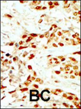
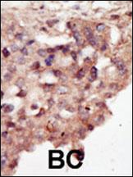
Formalin-fixed and paraffin-embedded human cancer tissue reacted with the primary antibody, which was peroxidase-conjugated to the secondary antibody, followed by DAB staining. This data demonstrates the use of this antibody for immunohistochemistry; clinical relevance has not been evaluated. BC = breast carcinoma; HC = hepatocarcinoma.
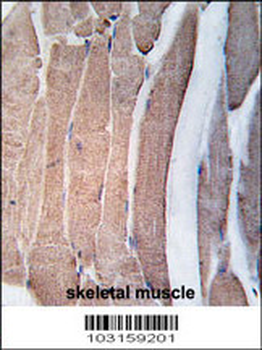
Formalin-fixed and paraffin-embedded human skeletal muscle tissue reacted with DAPK2 Antibody (N-term V55), which was peroxidase-conjugated to the secondary antibody, followed by DAB staining. This data demonstrates the use of this antibody for immunohistochemistry; clinical relevance has not been evaluated.
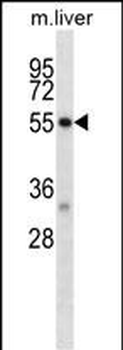
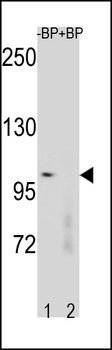
The anti-DAPK2 Pab is used in Western blot to detect DAPK2 in mouse lung tissue lysate.

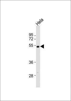
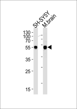
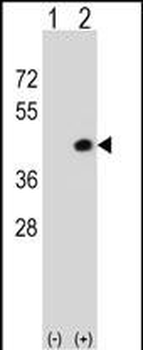
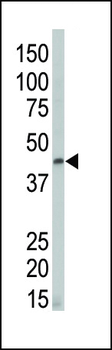
Western blot analysis of DAPK2 (arrow) using rabbit polyclonal DAPK2 Antibody (N-term). 293 cell lysates (2 ug/lane) either nontransfected (Lane 1) or transiently transfected with the DAPK2 gene (Lane 2).
Quick Database Links
UniProt Details
− No UniProt data available
NCBI Reference Sequences
−Associated Accession Numbers
Curated reference sequences for the gene transcript and protein product| Protein | NP_055141.2 |
|---|
Documents Download
Datasheet
Product Information
Request a Document
Protocol Information
WB
Western Blot (IB, immunoblot)
IHC-P
Immunohistochemistry Paraffin
DAPK2 Antibody (N-term) (orb1429149)

- 0.0
Based on 0 reviews
Participating in our Biorbyt product reviews program enables you to support fellow scientists by sharing your firsthand experience with our products.
Login to Submit a ReviewAvailable Sizes
Select a size below
Choose Conjugation or Carrier Free Version
Free Secondary Antibody (20 ul)0/0
Please add an antibody product to your cart first.