You have no items in your shopping cart.
Featured
KO/KD
Validated
Validated
Description
Research Area
Immunology & Inflammation
Images & Validation
−Item 1 of 12
| Tested Applications | ELISA, FC, ICC, IF, IHC-P, KO/KD Validated, WB |
|---|---|
| Reactivity | Human, Mouse, Rat |
| Predicted Reactivity | Bovine, Porcine |
Key Properties
−| Antibody Type | Primary Antibody |
|---|---|
| Host | Rabbit |
| Clonality | Polyclonal |
| Isotype | IgG |
| Immunogen | Anti-CXCR4 antibody (orb1239316) was raised against a peptide corresponding to 14 amino acids near the amino terminus of human CXCR4 isoform b. The immunogen is located within the first 50 amino acids of CXCR4. |
| Target | CXCR4 |
| Molecular Weight | Predicted: 40 kDa Observed: 44 kDa (Post-modification: 2 N-linked glycosylation) |
| Purification | CXCR4 Antibody is affinity chromatography purified via peptide column. |
| Conjugation | Unconjugated |
Storage & Handling
−| Storage | Maintain refrigerated at 2-8°C for up to 2 weeks. For long term storage store at -20°C in small aliquots to prevent freeze-thaw cycles. |
|---|---|
| Form/Appearance | Liquid |
| Buffer/Preservatives | CXCR4 Antibody is supplied in PBS containing 0.02% sodium azide. |
| Concentration | 1 mg/mL |
| Expiration Date | 12 months from date of receipt. |
| Disclaimer | For research use only |
Alternative Names
−CXCR4 Antibody: FB22, HM89, LAP3, LCR1, NPYR, WHIM, CD184, LESTR, NPY3R, NPYRL, HSY3RR, NPYY3R, D2S201E
Similar Products
−- Item 1 of 8
CD184/CXCR4 Rabbit Polyclonal Antibody [orb381896]
ICC, IF, IHC-P
Guinea pig, Human, Mouse, Rat
Rabbit
Polyclonal
Unconjugated
100 μg - Item 1 of 9
CXCR4 Rabbit Polyclonal Antibody [orb573686]
FC, IHC, WB
Equine, Porcine, Rabbit
Human, Mouse
Rabbit
Polyclonal
Unconjugated
100 μl - Item 1 of 6
CXCR4 Rabbit Polyclonal Antibody [orb10305]
IF, IHC-Fr, IHC-P, WB
Bovine, Rabbit
Human, Mouse, Rat
Rabbit
Polyclonal
Unconjugated
50 μl, 100 μl, 200 μl - Item 1 of 9
CXCR4 Antibody [orb1239315]
ELISA, IF, IHC, KO/KD Validated, WB
Bovine, Porcine, Sheep
Human, Mouse, Rat
Rabbit
Polyclonal
Unconjugated
0.02 mg, 0.1 mg - Item 1 of 5


Quality Guarantee
Explore bioreagents carefree to elevate your research. All our products are rigorously tested for performance. If a product does not perform as described on its datasheet, our scientific support team will provide expert troubleshooting, a prompt replacement, or a refund. For full details, please see our Terms & Conditions and Buying Guide. Contact us at [email protected].
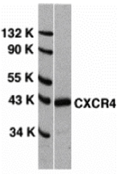
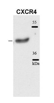
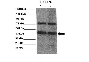
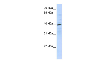
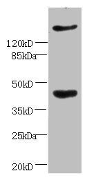
Western Blot Validation of CXCR4 in HeLa Cells, Loading: 15 µg of lysates per lane. Antibodies: orb1239316 (1 µg/mL), 1 h incubation at RT in 5% NFDM/TBST. Secondary: Goat anti-rabbit IgG HRP conjugate at 1:10000 dilution.
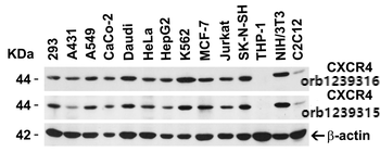
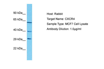
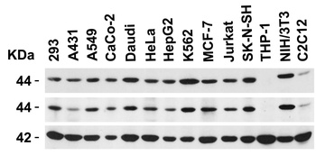
Independent Antibody Validation (IAV) via Protein Expression Profile in Cell Lines, Loading: 15 µg of lysates per lane. Antibodies: orb1239316 (1 µg/mL), orb1239315 (1 µg/mL), and beta-actin (1 µg/mL), 1 h incubation at RT in 5% NFDM/TBST. Secondary: Goat anti-rabbit IgG HRP conjugate at 1:10000 dilution.
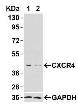
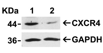
Validation with CXCR4 siRNA Knockdown in HeLa Cells, HeLa cells were transfected with control siRNAs (lane 1) or CXCR4 siRNAs (lane 2) Loading: 10 µg of HeLa whole cell lysates per lane. Antibodies: orb1239316 (2 µg/mL), 1 h incubation at RT in 5% NFDM/TBST. Secondary: Goat anti-rabbit IgG HRP conjugate at 1:10000 dilution.
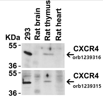
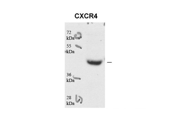
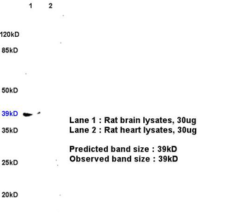
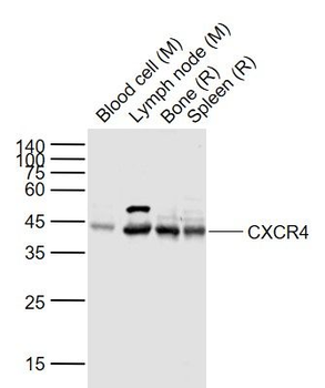
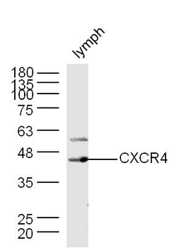
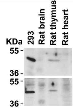
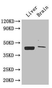
Animal Species Reactivity, Loading: Lysates/proteins at 20 µg per lane. Antibodies: orb1239316 (2 µg/mL) or orb1239315 (2 µg/mL). 1 h incubation at RT in 5% NFDM/TBST. Secondary: Goat anti-rabbit IgG HRP conjugate at 1:10000 dilution.
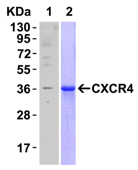
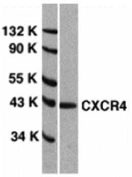
Recombinant Protein Test, Loading: CXCR4 partial recombinant protein. Lane 1: Anti-CXCR4 antibody (0.1 µg/mL) 1 h incubation at RT in 5% NFDM/TBST. Lane 2: Coomassie blue staining. Secondary: Goat anti-rabbit IgG HRP conjugate at 1:10000 dilution.
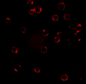
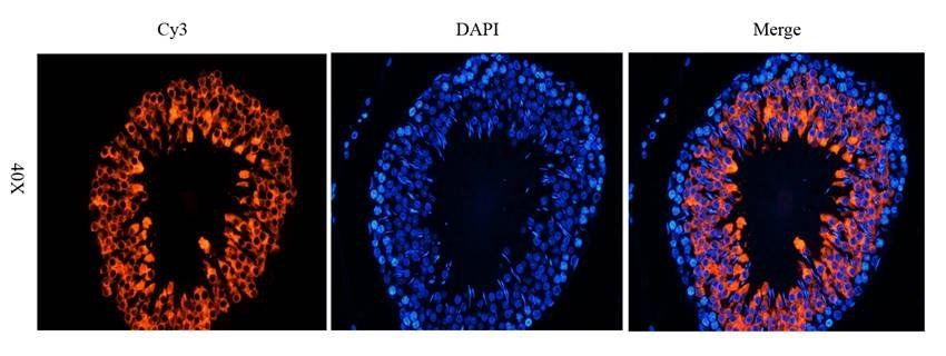
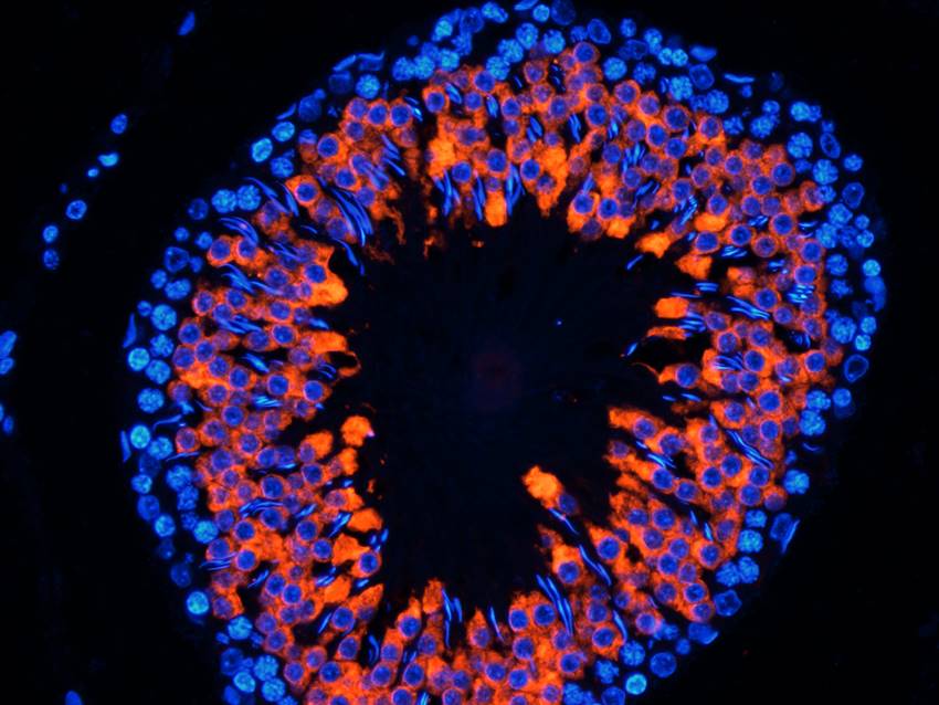
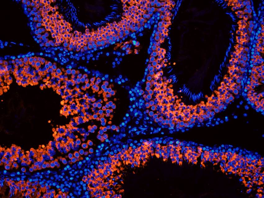
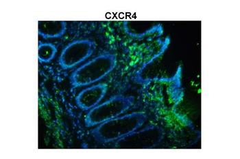
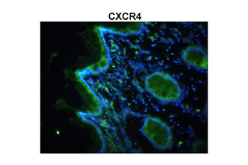
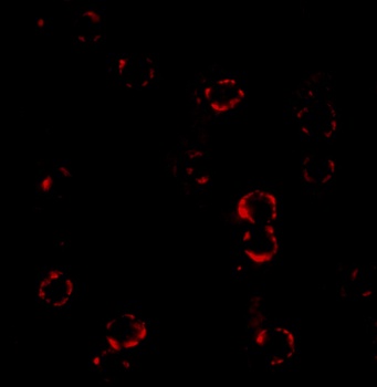
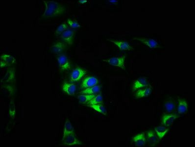
Immunofluorescence Validation of CXCR4 in HeLa Cells, Immunofluorescent analysis of 4% paraformaldehyde-fixed HeLa cells labeling CXCR4 with orb1239316 at 20 µg/mL, followed by goat anti-rabbit IgG secondary antibody at 1/500 dilution (red). Image showing both membrane and cytoplasmic staining on HeLa cells.
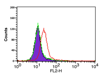
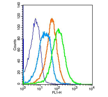
Flow Cytometry Validation of CXCR4 in HeLa Cells, Overlay histogram showing HeLa cells stained with orb1239316 (red line, 1 µg/1x106 cells). 1 h incubation at 4°C in 2% FBS/PBS. Followed by secondary antibody 488 goat anti-rabbit IgG (H+L) at 1/500 dilution for 1 h 4°C. Isotype control antibody (Green line) was mouse IgG1 (1 µg/1x106 cells) used under the same conditions.
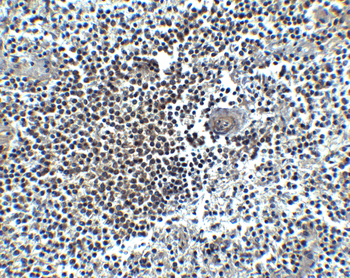
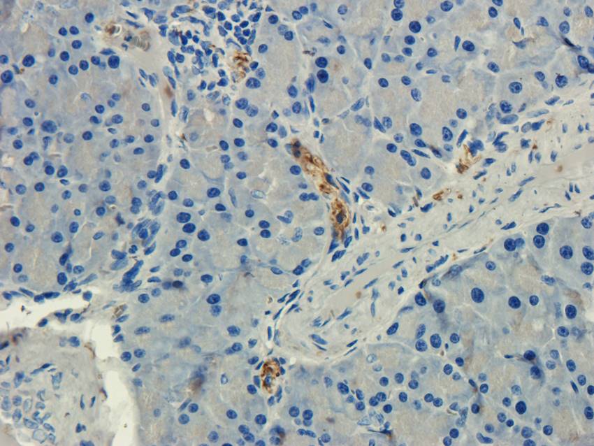
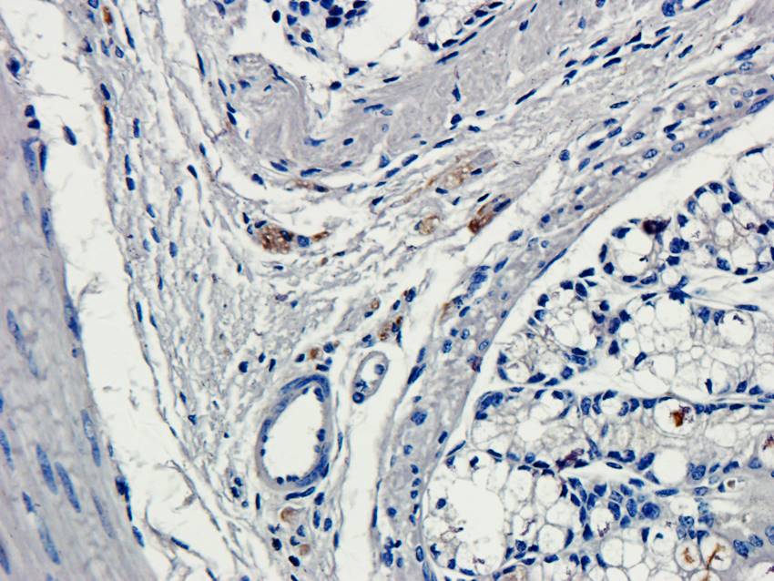
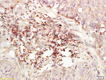
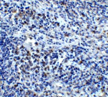
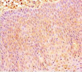
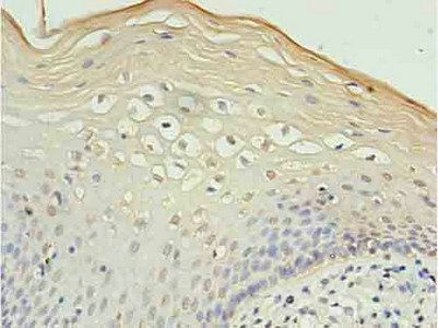
Immunohistochemistry Validation of CXCR4 in Human Spleen, Immunohistochemical analysis of paraffin-embedded human spleen tissue using anti-CXCR4 antibody (orb1239316) at 5 µg/mL. Tissue was fixed with formaldehyde and blocked with 10% serum for 1 h at RT; antigen retrieval was by heat mediation with a citrate buffer (pH6). Samples were incubated with primary antibody overnight at 4°C. A Goat anti-rabbit IgG H&L (HRP) at 1/250 was used as secondary. Counter stained with Hematoxylin.
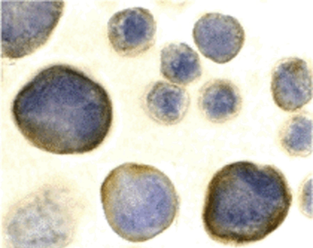
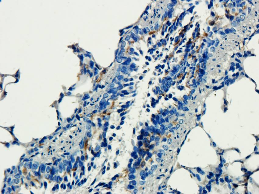
Immunocytochemistry Validation of CXCR4 in HeLa Cells, Immunocytochemical analysis of HeLa cells using anti-CXCR4 antibody (orb1239316) at 2 µg/mL. Cells was fixed with formaldehyde and blocked with 10% serum for 1 h at RT; antigen retrieval was by heat mediation with a citrate buffer (pH6). Samples were incubated with primary antibody overnight at 4°C. A goat anti-rabbit IgG H&L (HRP) at 1/250 was used as secondary. Counter stained with Hematoxylin.
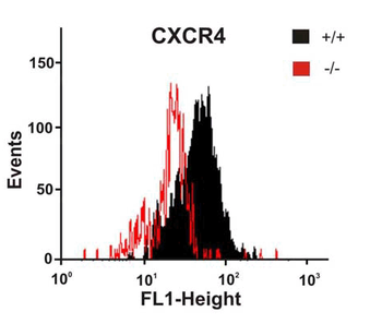
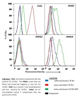
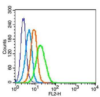
KO Validation of CXCR4 by Flow Cytometry, Astrocytes from wild-type or CXCR4 knockout mice were stained with primary antibodies against CXCR4 and FITC-labeled secondary antibodies, and subsequently subjected to flow cytometry. CXCR4−/− astrocytes (red) showed loss of CXCR4 cell-surface expression compared with wild-type cells (black).
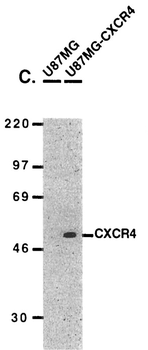
Overexpression Validation of CXCR4, U87MG and U87MG-CXCR4 extracts were included as negative and positive controls, respectively, for CXCR4 detection with anti-CXCR4 antibodies.

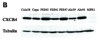
WB Validation of CXCR4 in Human Metastatic Melanoma, CXCR4 protein was detected in the human metastatic melanoma cell lines and human melanoma cell line (colo38), but not in the human primary melanocytes (MPR1) with anti-CXCR4 antibodies.
Documents Download
Datasheet
Product Information
Request a Document
Protocol Information
WB
Western Blot (IB, immunoblot)
IHC-P
Immunohistochemistry Paraffin
FC
Flow Cytometry
IF
Immunofluorescence
ICC
Immunocytochemistry
ELISA
Enzyme-linked Immunosorbent Assay (EIA)
CXCR4 Antibody (orb1239316)

- 0.0
Based on 0 reviews
Participating in our Biorbyt product reviews program enables you to support fellow scientists by sharing your firsthand experience with our products.
Login to Submit a ReviewAvailable Sizes
Select a size below
Free Secondary Antibody (20 ul)0/0
Please add an antibody product to your cart first.