
You have no items in your shopping cart.
Fluorescent Protein Research
Clover Fluorescent Protein: Bright Green FRET Donor and mClover Antibody
Explore the fluorescence properties of Clover fluorescent protein and mClover, compare Clover with EGFP and mClover3, understand its role in FRET biosensors, and detect mClover-tagged proteins with Biorbyt reagents.
1. What Is Clover Fluorescent Protein?
Clover fluorescent protein is an engineered green fluorescent protein derived from Aequorea victoria GFP. It was developed as a bright, efficiently folding fluorescent protein for live-cell imaging and genetically encoded Förster resonance energy transfer (FRET) biosensors.
Clover has excitation and emission maxima of approximately 505 nm and 515 nm, positioning its spectrum between EGFP and yellow fluorescent proteins. Its high extinction coefficient and quantum yield produce a strong green signal and make Clover an effective donor for red fluorescent protein acceptors.
The terms Clover, mClover and mClover3 refer to related but not identical constructs. Researchers should verify the exact sequence, monomerizing mutations and fluorescent-protein generation used in their plasmid or fusion protein.
Key point: Clover is best known for its high molecular brightness and its use as a green FRET donor. The later mClover3 variant was engineered to improve monomeric behaviour and photostability for demanding live-cell experiments.
Clover at a Glance
| Property | Clover |
|---|---|
| Protein family | Engineered Aequorea victoria GFP |
| Fluorescence colour | Bright green |
| Excitation maximum | Approximately 505 nm |
| Emission maximum | Approximately 515 nm |
| Approximate molecular weight | Approximately 27 kDa |
| Primary advantage | High molecular brightness and strong FRET donor performance |
| Common applications | FRET biosensors, fusion-protein imaging, reporter assays and protein localization |
| Detection methods | Direct fluorescence and anti-mClover antibody detection |
2. Clover vs EGFP and mClover3
Clover, EGFP and mClover3 all produce green fluorescence, but they were developed for different experimental priorities. EGFP remains a widely used general-purpose reporter, while Clover was optimized for brightness and FRET. mClover3 is a later monomeric generation designed for improved photostability and fusion-protein performance.
| Feature | EGFP | Clover | mClover3 |
|---|---|---|---|
| Excitation / emission | Approximately 488 / 507 nm | Approximately 505 / 515 nm | Approximately 506 / 518 nm |
| Main design priority | General imaging and reporter expression | High brightness and FRET performance | Improved monomeric and photostable performance |
| Typical FRET partner | Various red or yellow acceptors | mRuby2 | mRuby3 |
| Routine fusion imaging | Widely used | Suitable, depending on construct | Preferred when strict monomeric behaviour is important |
| Best selected when | A standard green reporter is required | Bright FRET donor performance is required | Long-term imaging and monomeric fusion performance are priorities |
Selection guidance: Use EGFP for routine green reporter applications, Clover for established Clover–mRuby2 FRET systems, and mClover3 when a newer monomeric and more photostable green FRET donor is required.
For a broader comparison of green fluorescent proteins, visit the Biorbyt GFP and EGFP resource page .
3. mClover Antibody for Fluorescent Fusion-Protein Detection
Biorbyt offers a directly relevant mClover antibody for detecting Clover fluorescent protein and mClover-tagged fusion proteins in Western blotting, immunofluorescence and immunohistochemistry workflows.
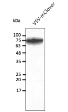
Western blot validation of mClover Antibody orb1463265 .
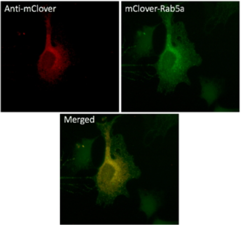
Immunofluorescence validation of mClover Antibody orb1463265 .
Direct Clover Detection Reagent
View orb1463265
mClover Antibody — orb1463265
orb1463265 is a goat polyclonal antibody developed for detection of mClover fluorescent protein. It is suitable for confirming expression, localization and molecular weight of mClover-tagged fusion proteins.
In product validation, the antibody detects an approximately 27 kDa band in transfected HEK293 cell lysate by Western blot. It has also been used for immunofluorescence detection in NIH3T3 cells expressing mClover-Rab5a.
Host: Goat
Clonality: Polyclonal
Isotype: IgG
Conjugation: Unconjugated
Applications:
IEM, IF, IHC-Fr, IHC-P, WB
WB dilution:
1:500–1:5,000
IHC / IF dilution:
1:50–1:500
Available size:
100 µg
Specificity note: Product validation states that orb1463265 does not recognize red fluorescent protein. This supports its use in dual-colour experiments where mClover is combined with an RFP-family marker.
4. Key Applications of Clover and mClover
FRET Biosensors
Clover is widely used as a green donor in genetically encoded FRET reporters. Changes in the donor-to-acceptor emission ratio can report kinase activity, small GTPase activity, membrane voltage and protein conformational changes.
Fusion-Protein Localization
Clover or mClover can be fused to a protein of interest to monitor subcellular localization, trafficking and redistribution in living cells. Antibody staining can confirm localization after fixation.
Reporter Expression Validation
Direct fluorescence indicates that the fluorescent domain has matured, while Western blot detection confirms expression and helps determine whether the complete fusion protein has the expected molecular weight.
Fixed-Cell Signal Amplification
Anti-mClover antibody staining can amplify weak fluorescence in fixed cells and tissues, supporting co-localization studies and experiments in which native fluorescence is reduced by sample preparation.
5. Detecting and Validating Clover Constructs
Clover and mClover constructs can be analysed by direct green fluorescence or antibody-based detection. For Biorbyt workflows, orb1463265 supports confirmation of mClover-tagged protein expression in WB, IF, IHC-Fr, IHC-P and IEM applications.
Direct Clover Fluorescence
Direct fluorescence is suitable for live-cell imaging, real-time localization and dynamic biosensor measurements.
- Suitable for living cells
- No antibody incubation required
- Preserves spatial and temporal information
- Supports FRET ratio measurements
- Does not confirm molecular weight or fusion integrity
Anti-mClover Antibody Detection
When fluorescence is weak or confirmation is required after fixation, orb1463265 can verify expression and visualize mClover-tagged proteins independently of fluorophore intensity.
- Use orb1463265 to confirm expression by Western blot
- Use orb1463265 in IF or IHC workflows to enhance detection in fixed samples
- Detect full-length and potentially degraded products
- Support co-localization with additional cellular markers
- Validate expression independently of fluorophore maturation
Essential Experimental Controls
- Untransfected or untagged sample: measures cellular autofluorescence and non-specific antibody binding.
- Known mClover-positive sample: confirms that the imaging system or orb1463265 detection workflow is functioning.
- Secondary-antibody-only control: measures background caused by the secondary antibody.
- Fusion-partner antibody or molecular-weight control: helps confirm that the complete fusion protein remains intact.
Interpretation: Green fluorescence confirms maturation of the Clover fluorescent domain, but it does not independently prove that the attached protein is intact, correctly localized or biologically active. Combining direct imaging with orb1463265 can provide stronger validation.
6. Clover in FRET Biosensors
Clover was originally developed as a bright green FRET donor. Its emission spectrum overlaps with the excitation spectrum of selected red fluorescent proteins while donor and acceptor emission remain sufficiently separated for ratiometric detection.
FRET Donor
Clover or mClover3
Green fluorescent protein excited near 505–506 nm
that transfers energy to a compatible red acceptor
when the proteins are sufficiently close.
→
FRET Acceptor
mRuby2 or mRuby3
Red fluorescent acceptor whose emission increases
when energy transfer occurs from the Clover-family
donor.
Why Use a Green–Red FRET Pair?
- Redder donor excitation can reduce cellular autofluorescence and phototoxicity compared with many cyan-donor systems.
- Clover–mRuby2 provides strong donor-emission and acceptor-excitation spectral overlap.
- Better separation between donor and acceptor emission can improve ratiometric signal interpretation.
- Clover-family pairs can be incorporated into kinase, GTPase, voltage and conformational biosensors.
FRET Design Considerations
- Confirm donor and acceptor orientation, linker length and fusion position.
- Include donor-only and acceptor-only controls for spectral bleed-through correction.
- Confirm that both fluorescent proteins mature efficiently in the selected cell type.
- Avoid assuming that high donor brightness alone will guarantee a large FRET response.
- Validate the reporter under the exact illumination, exposure and acquisition settings used in the final experiment.
Detection note: orb1463265 is intended for mClover detection. It should not be described as an mRuby2 or mRuby3 detection reagent, and recognition of newer Clover variants should be confirmed separately.
7. Companion Reagents and Related Resources
Clover experiments may require additional fluorescent protein information, secondary antibodies, Western blot detection reagents and red fluorescent protein resources.
Green Fluorescent Proteins
GFP and EGFP Resource Page
Compare GFP-family fluorescent proteins and explore
Biorbyt antibodies, VHH single-domain antibodies and
recombinant proteins.
Research Hub
Fluorescent Protein Hub
Explore green, yellow, red and far-red fluorescent
proteins for imaging, reporter and biosensor
applications.
Red Fluorescent Proteins
mCherry Fluorescent Protein Resource
Explore red fluorescent protein imaging and
antibody-detection workflows for dual-colour
experiments.
Western Blot Detection
ECL Plus Substrate — orb527071
Chemiluminescent HRP substrate for sensitive Western
blot detection of mClover-tagged fusion proteins.
Custom Service
Antibody Conjugation
Explore custom fluorophore, enzyme and biotin
conjugation options for compatible Biorbyt
antibodies.
8. Why Choose Biorbyt for mClover Detection?
Direct mClover Product
orb1463265 is specifically described as an antibody to mClover fluorescent protein.
Multiple Validated Applications
The antibody supports WB, IF, IHC-Fr, IHC-P and IEM workflows.
Imaging and WB Validation
Product data include transfected-cell Western blot and cellular immunofluorescence validation.
RFP Non-Reactivity Data
Product information states that the antibody does not recognize red fluorescent protein.
9. Frequently Asked Questions
What is Clover fluorescent protein?
Clover is an engineered GFP-derived green fluorescent protein developed for high brightness, live-cell imaging and FRET biosensor applications. Its excitation and emission maxima are approximately 505 nm and 515 nm.
What is the difference between Clover and EGFP?
EGFP is a general-purpose green reporter with excitation and emission maxima near 488 nm and 507 nm. Clover is red-shifted and was optimized for high molecular brightness and FRET donor performance.
What is the difference between Clover and mClover3?
mClover3 is a later monomeric Clover-family variant engineered for improved photostability and fusion-protein performance. It is not identical in sequence to the original Clover construct.
Which FRET acceptor is commonly paired with Clover?
The classical Clover donor is commonly paired with mRuby2. The later mClover3 generation is commonly paired with mRuby3 in updated green–red FRET systems.
Can mClover be detected without an antibody?
Yes. Mature mClover can be detected directly through its green fluorescence. Antibody detection is useful for signal amplification, fixed-sample staining and molecular-weight confirmation.
Can orb1463265 detect mClover3?
The public product information identifies orb1463265 as an mClover antibody but does not explicitly state validation against mClover3. Compatibility should therefore be confirmed using a known positive mClover3 sample.
10. Scientific References
- Lam AJ, St-Pierre F, Gong Y, et al. Improving FRET dynamic range with bright green and red fluorescent proteins. Nature Methods. 2012;9:1005–1012. doi:10.1038/nmeth.2171
- Bajar BT, Wang ES, Lam AJ, et al. Improving brightness and photostability of green and red fluorescent proteins for live cell imaging and FRET reporting. Scientific Reports. 2016;6:20889. doi:10.1038/srep20889
- Cranfill PJ, Sell BR, Baird MA, et al. Quantitative assessment of fluorescent proteins. Nature Methods. 2016;13:557–562. doi:10.1038/nmeth.3891
- Campbell BC, Petsko GA, Liu CF. Crystal Structure of Green Fluorescent Protein Clover and Design of Clover-Based Redox Sensors. Structure. 2018;26:225–237.e3. doi:10.1016/j.str.2017.12.006
Scientifically reviewed by:
Biorbyt Scientific Support Team
Last reviewed:
July 2026