You have no items in your shopping cart.
CD99 Antibody
SKU: orb749672
Description
Research Area
Epigenetics
Images & Validation
−Item 1 of 1
| Tested Applications | IHC-P |
|---|---|
| Dilution Range | Immunohistochemistry (FFPE): 1-2ug/ml for 30 min at RT |
| Reactivity | Human |
| Application Notes |
Key Properties
−| Antibody Type | Primary Antibody |
|---|---|
| Host | Mouse |
| Clonality | Monoclonal |
| Isotype | Mouse IgG1, kappa |
| Clone No. | SPM596 |
| Immunogen | Recombinant full-length human protein was used as the immunogen for the anti-CD99 antibody. |
| Purification | Protein G affinity chromatography |
| Conjugation | Unconjugated |
Storage & Handling
−| Storage | Maintain refrigerated at 2-8°C for up to 2 weeks. For long term storage store at -20°C in small aliquots to prevent freeze-thaw cycles. |
|---|---|
| Buffer/Preservatives | 0.2 mg/ml in 1X PBS with 0.1 mg/ml rAlbumin and 0.05% sodium azide |
| Expiration Date | 12 months from date of receipt. |
| Disclaimer | For research use only |
Similar Products
−- Item 1 of 1
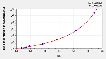
Human Cluster of Differentiation 99 (CD99) ELISA Kit [orb776723]
Human
0.16-10 ng/mL
0.059 ng/mL
96 T, 48 T - Item 1 of 1
Mouse Cluster of Differentiation 99 (CD99) ELISA Kit [orb779426]
Mouse
0.16-10 ng/mL
0.067 ng/mL
48 T, 96 T - Item 1 of 3
CD99 Antibody [orb388433]
FC, IF, IHC
Human
Mouse
Monoclonal
Unconjugated
20 μg, 100 μg, 100 μg (without BSA and Azide) - Item 1 of 3
- Item 1 of 2
CD99 Antibody [orb1410584]
IHC
Human
Mouse
Monoclonal
Unconjugated
20 μg, 100 μg, 100 μg (without BSA and Azide)


Quality Guarantee
Explore bioreagents carefree to elevate your research. All our products are rigorously tested for performance. If a product does not perform as described on its datasheet, our scientific support team will provide expert troubleshooting, a prompt replacement, or a refund. For full details, please see our Terms & Conditions and Buying Guide. Contact us at [email protected].
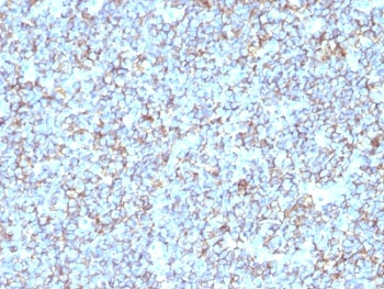
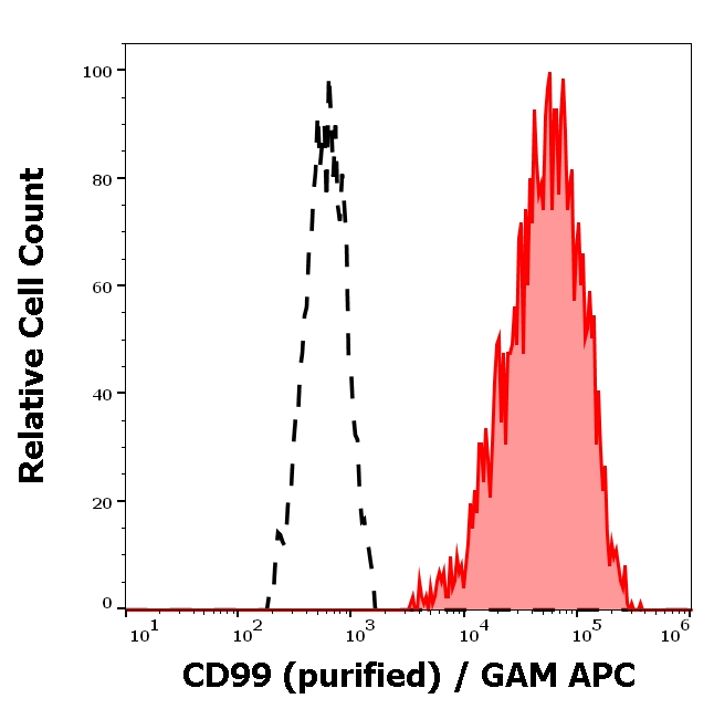
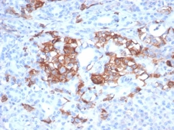
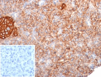
IHC: Formalin-fixed, paraffin-embedded human Ewing's sarcoma stained with anti-CD99 antibody (SPM596).
Quick Database Links
UniProt
UniProt Details
− No UniProt data available
Documents Download
Datasheet
Product Information
Request a Document
CD99 Antibody (orb749672)

- 0.0
Based on 0 reviews
Participating in our Biorbyt product reviews program enables you to support fellow scientists by sharing your firsthand experience with our products.
Login to Submit a ReviewAvailable Sizes
Select a size below