You have no items in your shopping cart.
KO/KD
Validated
Validated
Description
Research Area
Immunology & Inflammation
Images & Validation
−Item 1 of 2
| Tested Applications | ELISA, IF, KO/KD Validated, WB |
|---|---|
| Dilution Range | ELISA: 1:10,000 - 1:50,000, IF: 1:1,000 - 1:5,000, WB: 1:1,000 - 1:5,000 |
| Reactivity | Human, Primate |
| Application Notes |
Key Properties
−| Antibody Type | Primary Antibody |
|---|---|
| Host | Rabbit |
| Clonality | Polyclonal |
| Isotype | IgG |
| Immunogen | This affinity purified antibody was prepared from whole rabbit serum produced by repeated immunizations with a recombinant protein corresponding to amino acids 1-512 (extracellular domain) of mouse CD97 protein. |
| Target | Cd97 |
| Purity | This Protein A purified antibody is directed against the mouse CD97 protein. The product was protein A purified from monospecific antiserum. A BLAST analysis was used to suggest cross reactivity with CD97 proteins from mouse (100%) and rat (74%). Only 49% homology is noted for the human homologue. Reactivity against CD97 from other sources is not known. |
| Conjugation | Unconjugated |
Storage & Handling
−| Storage | Store vial at 4° C prior to restoration. For extended storage aliquot contents and freeze at -20° C or below. Avoid cycles of freezing and thawing. Centrifuge product if not completely clear after standing at room temperature. This product is stable for several weeks at 4° C as an undiluted liquid. Dilute only prior to immediate use. |
|---|---|
| Form/Appearance | Lyophilized |
| Buffer/Preservatives | Preservative: 0.01% (w/v) Sodium Azide. Stabilizer: None; Buffer: 0.02 M Potassium Phosphate, 0.15 M Sodium Chloride, pH 7.2 |
| Concentration | 5.0 mg/mL |
| Expiration Date | 12 months from date of receipt. |
| Disclaimer | For research use only |
Alternative Names
−rabbit anti-CD97 Antibody, CD97 antigen antibody, CD97 molecule antibody, Leukocyte antigen CD97 antibody, CD97 antigen subunit alpha, CD97 antigen subunit beta
Similar Products
−- Item 1 of 1
- Item 1 of 3
CD97/Adgre5 Rabbit Polyclonal Antibody [orb738372]
ELISA, FC, IHC, WB
Mouse, Rat
Rabbit
Polyclonal
Unconjugated
100 μg - Item 1 of 2
Goat anti-Cd97 (mouse) Antibody [orb167690]
ELISA, KO/KD Validated, WB
Mouse
Polyclonal
Unconjugated
100 μg - Item 1 of 2
- Item 1 of 2

Quality Guarantee
Explore bioreagents carefree to elevate your research. All our products are rigorously tested for performance. If a product does not perform as described on its datasheet, our scientific support team will provide expert troubleshooting, a prompt replacement, or a refund. For full details, please see our Terms & Conditions and Buying Guide. Contact us at [email protected].
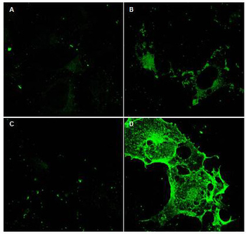
Immunofluorescence Microscopy using Biorbyt's Protein A purified anti-CD97 antibody shows staining of Fc-CD97-(5EGF) (panel D) in transfected COS cells. Panel A and C shows similar staining using pre-immune serum. Panel A and B show staining of mock transfected COS cells (no vector). A 1:2500 dilution of the primary antibody was used.
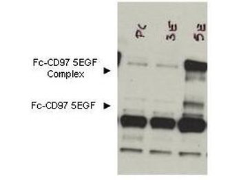
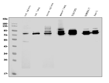
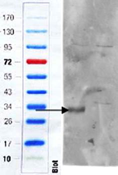
Western blot using Biorbyt's Protein A purified anti-CD97 antibody Lane 1: lysate from cells transfected with control DNA. Lane 2: bone marrow lysates taken from CD97 knockout mice. Lane 3: lysate from COS cells expressing Fc-CD97- (5EGF). Load: 10 µl per lane. Primary Antibody: 1:1000 dilution of the primary antibody was used. Secondary Antibody: Exposure: 10-sec. Results: The formation of the CD97 complex is currently under investigation. A shows detection of bands corresponding to free Fc-CD97- (5EGF) (lower arrowhead) and Fc-CD97- (5EGF) present as a complex (upper arrowhead) in lysates from COS cells. No staining was noted from bone marrow lysates taken from CD97 knockout mice. ~65 kDa appearing in all lanes is not known.
Documents Download
Datasheet
Product Information
Request a Document
Protocol Information
WB
Western Blot (IB, immunoblot)
IF
Immunofluorescence
ELISA
Enzyme-linked Immunosorbent Assay (EIA)
Cd97 Antibody (orb344630)

- 0.0
Based on 0 reviews
Participating in our Biorbyt product reviews program enables you to support fellow scientists by sharing your firsthand experience with our products.
Login to Submit a ReviewAvailable Sizes
Select a size below
Free Secondary Antibody (20 ul)0/0
Please add an antibody product to your cart first.