You have no items in your shopping cart.
Featured
Description
Research Area
Immunology & Inflammation
Images & Validation
−Item 1 of 10
| Tested Applications | ELISA, ICC, IF, IHC-P, WB |
|---|---|
| Dilution Range | WB: 1:200-2000, IF/ICC: 1:50-300, IHC-P: 1:100-500 (based on 0.5 mg/ml) |
| Reactivity | Human, Mouse, Rat |
Key Properties
−| Host | Rabbit |
|---|---|
| Clonality | Polyclonal |
| Isotype | IgG |
| Immunogen | KLH conjugated synthetic peptide derived from human CD19. Please contact us for the exact immunogen sequence. The peptide is available as orb374859. |
| Target | CD19 |
| Molecular Weight | 128 kDa |
| Purity | Polyclonal antibodies are purified by peptide affinity chromatography |
| Conjugation | Unconjugated |
Storage & Handling
−| Storage | Maintain refrigerated at 2-8°C for up to 2 weeks. For long term storage store at -20°C in small aliquots to prevent freeze-thaw cycles. |
|---|---|
| Form/Appearance | 10 mM PBS, 0.02% sodium azide |
| Concentration | - 100 μg (in 200 μl): 0.5 mg/ml- 200 μg (in 400 μl): 0.5 mg/ml |
| Expiration Date | 12 months from date of receipt. |
| Disclaimer | For research use only |
Alternative Names
−anti-Cluster Of Differentiation 19 antibody, anti-AW495831 antibody, anti-B lymphocytegen CD19 antibody, anti-B lymphocyte surfacegen B4 antibody, anti-B4 antibody, anti-CD19 antibody, anti-CD19gen antibody, anti-CD19 molecule antibody, anti-Cd19 proteinbod antibody, anti-deficiency due to defect in CD19 antibody, anti-Differentiationgen CD19 antibody, anti-included antibody, anti-LEU-12 antibody, anti-Lymphocyte Surfacegen antibody, anti-MGC12802 antibody
Similar Products
−- Item 1 of 6
CD19 Rabbit Polyclonal Antibody [orb251482]
ELISA, ICC, IF, IHC-P
Bovine, Equine, Guinea pig, Human, Mouse, Porcine, Rat
Rabbit
Polyclonal
Unconjugated
100 μg - Item 1 of 6
CD19 Antibody (N-term) [orb1935338]
FC, IF, IHC-P, WB
Human
Rabbit
Polyclonal
Unconjugated
50 μl, 100 μl - Item 1 of 3
CD19 Rabbit Polyclonal Antibody [orb317567]
FC, IF, IHC-Fr, IHC-P
Bovine, Equine, Guinea pig, Porcine, Rat
Human, Mouse
Rabbit
Polyclonal
Unconjugated
50 μl, 100 μl, 200 μl - Item 1 of 4
CD19 rabbit pAb Antibody [orb767185]
ELISA, FC, IF, IHC, WB
Human, Mouse, Rat
Polyclonal
Unconjugated
100 μl - Item 1 of 1
CD19 Rabbit Polyclonal Antibody [orb1699]
FC
Guinea pig, Mouse, Porcine, Rat
Human
Rabbit
Polyclonal
Unconjugated
50 μl, 100 μl, 200 μl


Quality Guarantee
Explore bioreagents carefree to elevate your research. All our products are rigorously tested for performance. If a product does not perform as described on its datasheet, our scientific support team will provide expert troubleshooting, a prompt replacement, or a refund. For full details, please see our Terms & Conditions and Buying Guide. Contact us at [email protected].
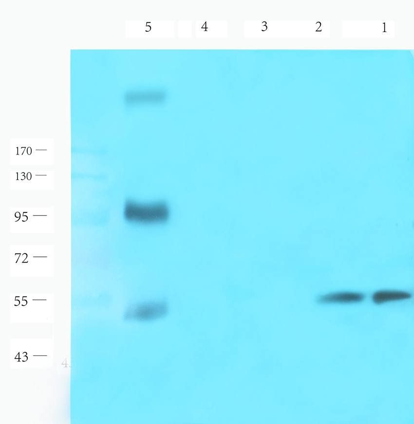
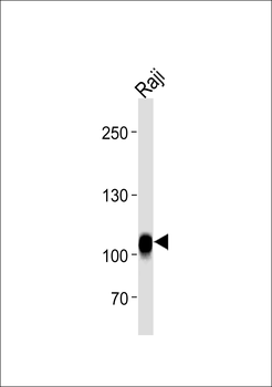
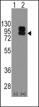
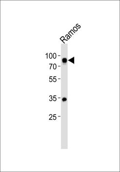
Western blot analysis of rat lymph node (lane 1), rat spleen (lane 2), rat lung (lane 3), mouse brain (lane 4), human breast cancer (lane 5), human ovarian cancer (lane 6) using CD19 antibody (1 ug/ml)
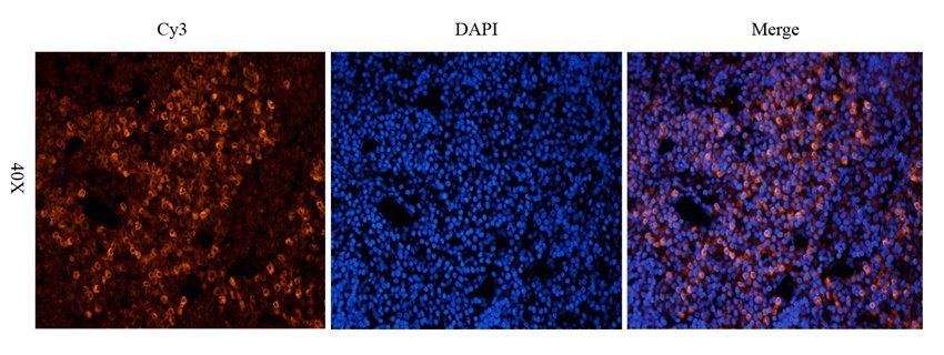
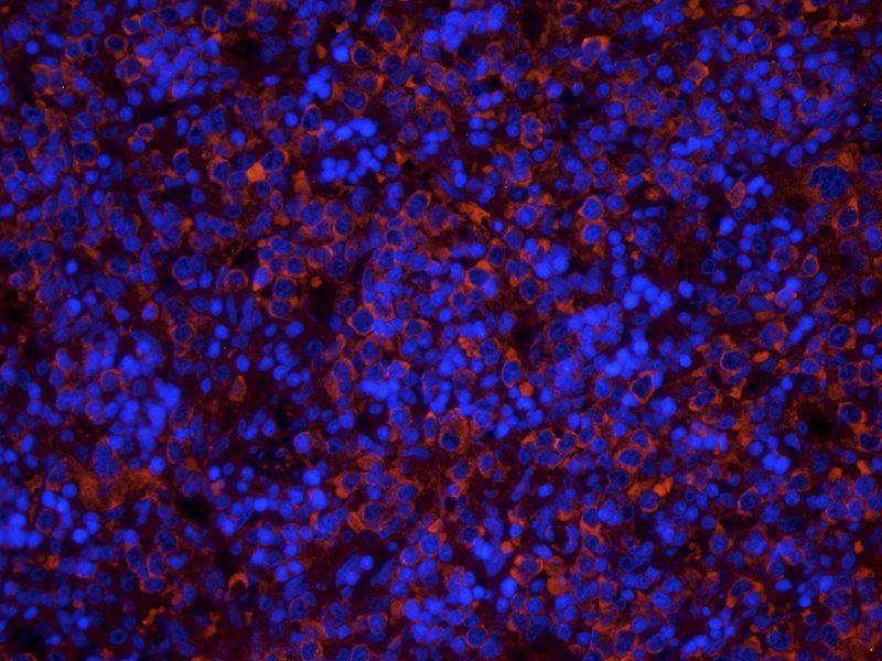
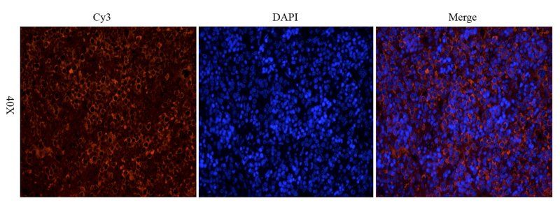
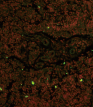
Immunofluorescence analysis of rat lymph node tissue using anti-CD19 (2.5 ug/ml)
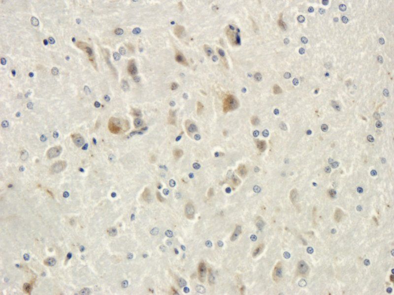
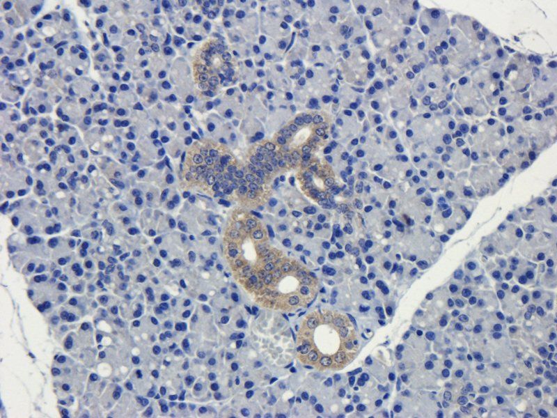
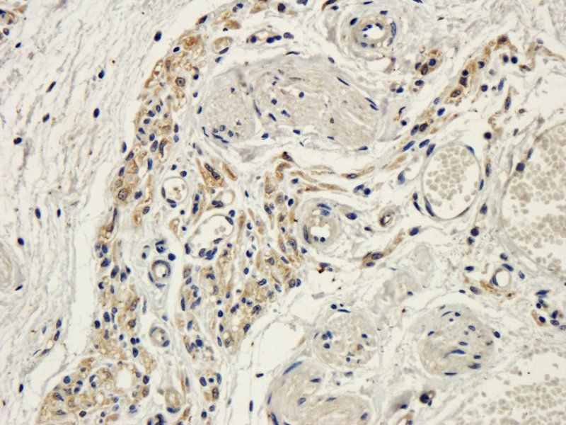
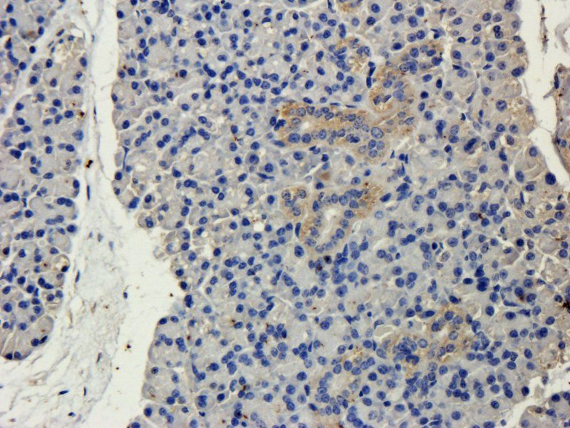
IHC-P staining of rat brain tissue using CD19 antibody (dilution at 2.5 ug/ml)
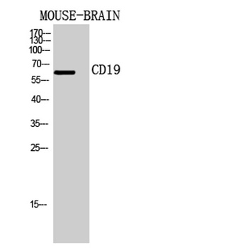
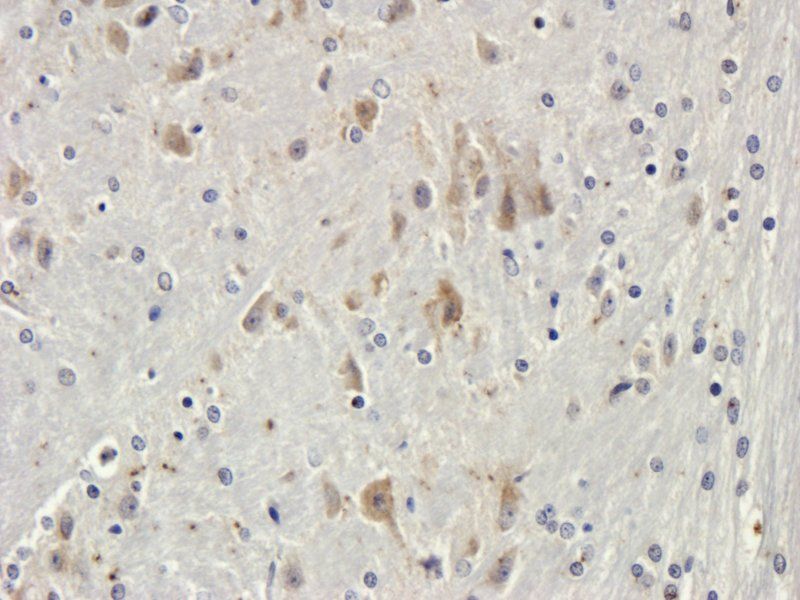
IHC-P staining of mouse brain tissue using anti-CD19 (dilution at 2.5 ug/ml)
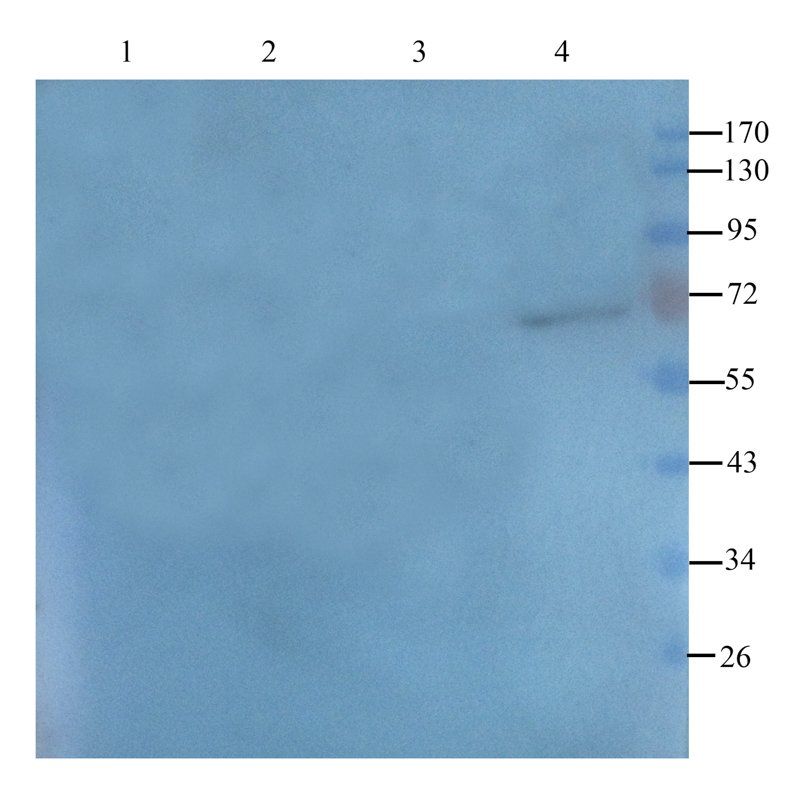
WB analysis of mouse lymph node (lane 1), mouse spleen (lane 2), mouse brain (lane 3); mouse liver (lane 4) using CD19 antbody (1 ug/ml)
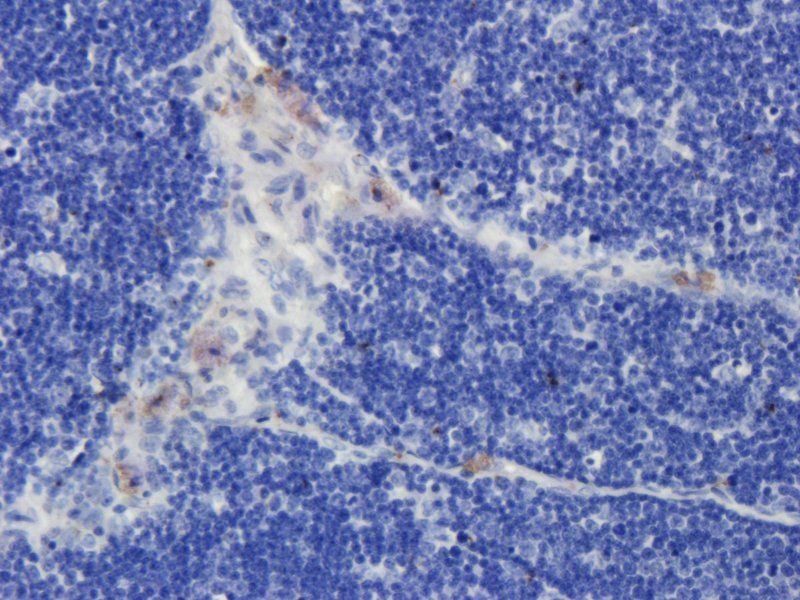
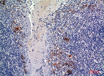
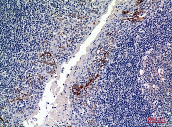
IHC-P image of rat thymus tissue using CD19 antibody (dilution of primary antibody at 2.5 ug/ml)
IHC-P staining of rat brain tissue using anti-CD19 (dilution at 2.5 ug/ml)
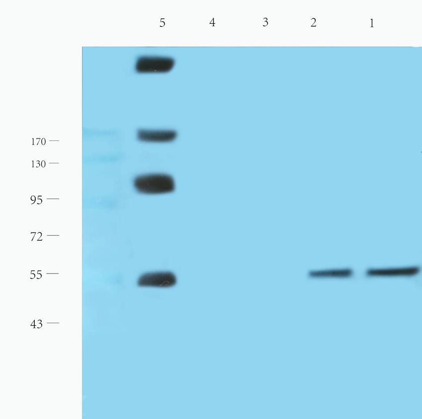
WB analysis of rat lymph node (lane 1), rat spleen (lane 2), rat lung (lane 3), mouse brain (lane 4), human breast cancer (lane 5), human ovarian cancer (lane 6) using CD19 antibody (1 ug/ml)
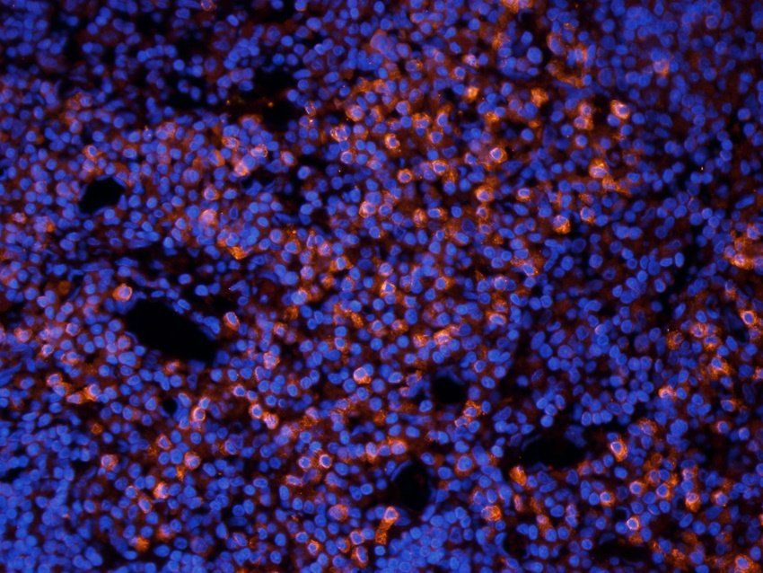
IF image of rat lymph node tissue using CD19 antibody (2.5 ug/ml)
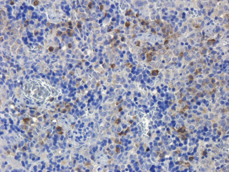
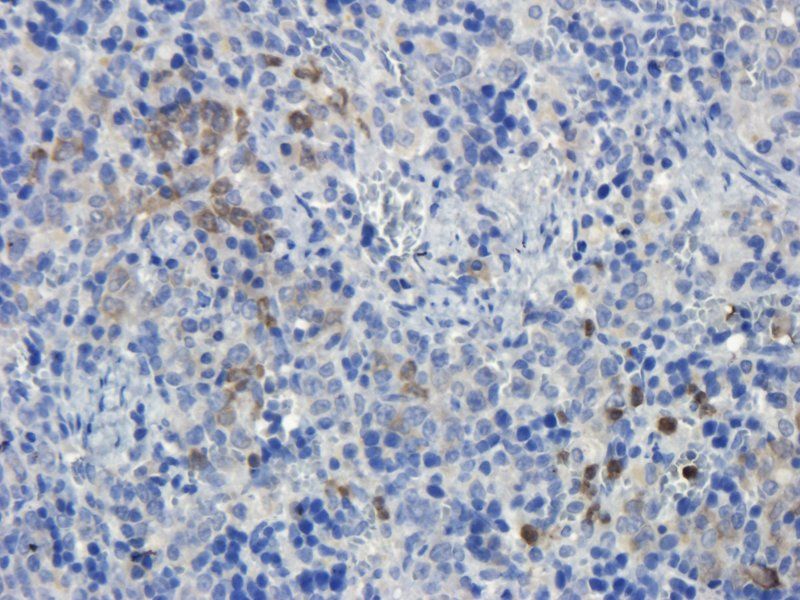
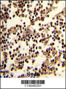
Immunohistochemical staining of paraffin embedded mouse lymph node tissue using anti-CD19 (primary antibody at 2.5 ug/ml)
Quick Database Links
Gene Symbol
CD19
UniProt
RefSeq (mRNA):NM_001770.5, NM_001770.51
RefSeq (Protein):NP_001761.31, NP_001761.3
UniProt Details
− No UniProt data available
NCBI Gene Details
− No NCBI Gene data available
NCBI Reference Sequences
−Associated Accession Numbers
Curated reference sequences for the gene transcript and protein product| mRNA | NM_001770.5, NM_001770.51 |
|---|---|
| Protein | NP_001761.31, NP_001761.3 |
Documents Download
Datasheet
Product Information
Request a Document
Protocol Information
WB
Western Blot (IB, immunoblot)
IHC-P
Immunohistochemistry Paraffin
IF
Immunofluorescence
ICC
Immunocytochemistry
ELISA
Enzyme-linked Immunosorbent Assay (EIA)
CD19 Rabbit Polyclonal Antibody (orb10306)

- 0.0
Based on 0 reviews
Participating in our Biorbyt product reviews program enables you to support fellow scientists by sharing your firsthand experience with our products.
Login to Submit a ReviewAvailable Sizes
Select a size below
Choose Conjugation or Carrier Free Version
Free Secondary Antibody (20 ul)0/0
Please add an antibody product to your cart first.