You have no items in your shopping cart.
Description
Research Area
Neuroscience
Images & Validation
−Item 1 of 3
| Tested Applications | FC, IHC-P, WB |
|---|---|
| Dilution Range | WB - 1:1000, IHC-P - 1:25, FC - 1:25 |
| Reactivity | Human |
Key Properties
−| Host | Mouse |
|---|---|
| Clonality | Monoclonal |
| Isotype | IgG1,k |
| Clone No. | B3598EV021X46X07 |
| Immunogen | This CAPN1 antibody is generated from a mouse immunized with a KLH conjugated synthetic peptide between amino acids from the human region of human CAPN1. Antigen Region: 1-420 aa. |
| Target | CAPN1 (HGNC:1476) |
| Molecular Weight | 81890 Da |
| Conjugation | Unconjugated |
Storage & Handling
−| Storage | Maintain refrigerated at 2-8°C for up to 2 weeks. For long term storage store at -20°C in small aliquots to prevent freeze-thaw cycles |
|---|---|
| Form/Appearance | Purified monoclonal antibody supplied in PBS with 0.09% (W/V) sodium azide. This antibody is purified through a protein G column, followed by dialysis against PBS. |
| Expiration Date | 12 months from date of receipt. |
| Disclaimer | For research use only |
Alternative Names
−Calpain-1 catalytic subunit, Calcium-activated neutral proteinase 1, CANP 1, Calpain mu-type, Calpain-1 large subunit, Cell proliferation-inducing gene 30 protein, Micromolar-calpain, muCANP, CAPN1, CANPL1
Similar Products
−- Item 1 of 1
Human Calpain 1, Large Subunit (CAPN1) ELISA Kit [orb777725]
Human
1.57-100 ng/mL
0.57 ng/mL
48 T, 96 T - Item 1 of 1
Mouse Calpain 1, Large Subunit (CAPN1) ELISA Kit [orb776578]
Mouse
1.57-100 ng/mL
0.62 ng/mL
48 T, 96 T - Item 1 of 1
- Item 1 of 4
Calpain 1/CAPN1 Rabbit Polyclonal Antibody [orb251513]
FC, ICC, IF, IHC, WB
Human, Mouse, Rat
Rabbit
Polyclonal
Unconjugated
100 μg - Item 1 of 4


Quality Guarantee
Explore bioreagents carefree to elevate your research. All our products are rigorously tested for performance. If a product does not perform as described on its datasheet, our scientific support team will provide expert troubleshooting, a prompt replacement, or a refund. For full details, please see our Terms & Conditions and Buying Guide. Contact us at [email protected].
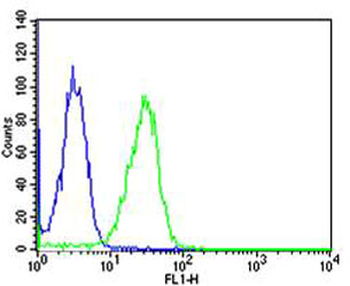
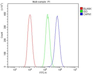
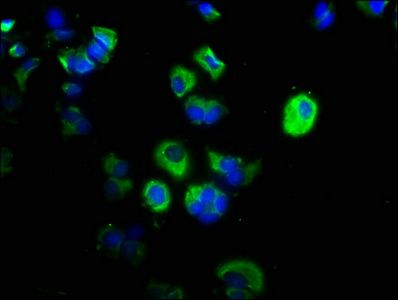
Flow cytometric analysis of Hela cells using CAPN1 Antibody (green) compared to an isotype control of mouse IgG1 (blue). Diluted at 1:25 dilution. An Alexa Fluor 488 goat anti-mouse lgG at 1:400 dilution was used as the secondary antibody.
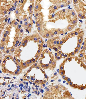
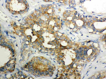
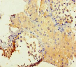
Immunohistochemical analysis of paraffin-embedded H. kidney section using CAPN1 Antibody. Diluted at 1:25 dilution. A peroxidase-conjugated goat anti-mouse IgG at 1:400 dilution was used as the secondary antibody, followed by DAB staining.
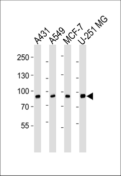
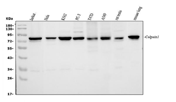
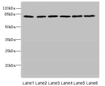
Western blot analysis of lysates from A431, A549, MCF-7, U-251 MG cell line (from left to right), using CAPN1 Antibody. Diluted at 1:1000 at each lane. A goat anti-mouse IgG H&L (HRP) at 1: 3000 dilution was used as the secondary antibody. Lysates at 35μg per lane.
Quick Database Links
Gene Symbol
CAPN1 (HGNC:1476)
UniProt
UniProt Details
− No UniProt data available
Documents Download
Datasheet
Product Information
Request a Document
Protocol Information
WB
Western Blot (IB, immunoblot)
IHC-P
Immunohistochemistry Paraffin
FC
Flow Cytometry
CAPN1 Antibody (orb1927151)

- 0.0
Based on 0 reviews
Participating in our Biorbyt product reviews program enables you to support fellow scientists by sharing your firsthand experience with our products.
Login to Submit a ReviewAvailable Sizes
Select a size below
Choose Conjugation or Carrier Free Version
Free Secondary Antibody (20 ul)0/0
Please add an antibody product to your cart first.