You have no items in your shopping cart.
Description
Images & Validation
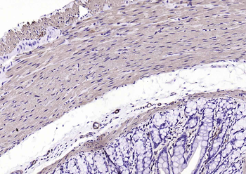
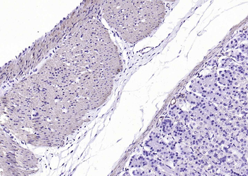
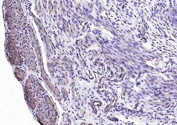
−| Tested Applications | ELISA, WB |
|---|---|
| Dilution Range | ELISA: 1:100,000, WB: 1:10,000 - 1:25,000 |
| Application Notes |
Key Properties
−| Antibody Type | Primary Antibody |
|---|---|
| Host | Rabbit |
| Clonality | Polyclonal |
| Isotype | Antiserum |
| Immunogen | Biotin conjugated to Keyhole Limpet Hemocyanin (KLH) |
| Purity | This product was prepared from monospecific antiserum by a delipidation and defibrination. Assay by immunoelectrophoresis resulted in a single precipitin arc against anti-rabbit serum, biotinylated IgG and Biotinylated BSA. |
| Conjugation | Unconjugated |
Storage & Handling
−| Storage | Store vial at 4° C prior to restoration. For extended storage aliquot contents and freeze at -20° C or below. Avoid cycles of freezing and thawing. Centrifuge product if not completely clear after standing at room temperature. This product is stable for several weeks at 4° C as an undiluted liquid. Dilute only prior to immediate use. |
|---|---|
| Form/Appearance | Lyophilized |
| Buffer/Preservatives | 0.01% (w/v) Sodium Azide |
| Concentration | 90 mg/mL |
| Expiration Date | 12 months from date of receipt. |
| Disclaimer | For research use only |
Alternative Names
−Similar Products
−Goat Anti-Rabbit IgG H&L, Biotin conjugated [orb868233]
ELISA, FC, ICC, IF, IHC-Fr, IHC-P, WB
Rabbit
Goat
Polyclonal
Biotin
100 μl, 1 mlGoat Anti-Mouse IgG H&L, Biotin conjugated [orb868227]
ELISA, FC, ICC, IF, IHC-Fr, IHC-P, WB
Mouse
Goat
Polyclonal
Biotin
100 μl, 1 mlF(ab')₂ Fragment Goat Anti-Mouse IgG H&L, Biotin conjugated [orb1974591]
ELISA, FC, ICC, IF, IHC-Fr, IHC-P, WB
Mouse
Goat
Polyclonal
Biotin
100 μlVPS35 Antibody (Biotin) [orb612801]
ICC, IHC, IP, KO/KD Validated, WB
Human, Mouse, Rat
Mouse
Monoclonal
Biotin
100 μg

Quality Guarantee
Explore bioreagents carefree to elevate your research. All our products are rigorously tested for performance. If a product does not perform as described on its datasheet, our scientific support team will provide expert troubleshooting, a prompt replacement, or a refund. For full details, please see our Terms & Conditions and Buying Guide. Contact us at [email protected].

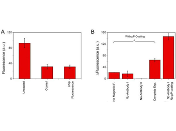
(a) Channels coating: remnant fluorescence due to nonspecific binding of anti-rabbit IgG labeled with B-rhodamine (p/n orb347647) [AbII] at a concentration of 100 µg/ml in uncoated and coated microchannels with silane-PEG. (b) Fluorescence obtained from the complete immunoassay at a flow rate of 5 µl/h and at a anti-biotin rabbit IgG (p/n orb750609) [AbI] concentration of 50 pg/ml is compared to the fluorescence obtained from the immunoassay performed without applying the magnetic field (column 1), without adding anti-biotin rabbit IgG (p/n orb750609) [AbI] (column 2), without adding), anti-rabbit IgG labeled with B-rhodamine (p/n orb347647) [AbII] (column 3), and, finally, the efficacy of the microparticles coating was tested by performing the immunoassay without anti-biotin rabbit IgG (p/n orb750609) [AbI] and with noncoated microparticles (column 5). The level of fluorescence of the two first columns in b results from nonspecific interactions of anti-rabbit IgG labeled with B-rhodamine (p/n orb347647) [AbII] with the microparticles. Error bars are standard deviation.

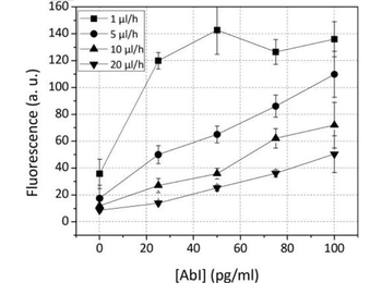
Shows the fluorescence difference from immunoassays at various concentrations of the primary antibody. Calibration curve: fluorescence difference from immunoassays with varying concentrations of anti-biotin rabbit IgG (p/n orb750609) [AbI] at different flow rates of the fluorogenic enzyme substrate anti-rabbit IgG labeled alkaline phosphatase was used (p/n orb347681). Each experiment was repeated on three different devices.

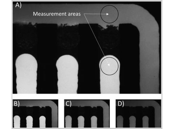
Typical experiment image where the complexes Ag–AbI–AbII are attached to the nanoparticles trapped in the magnetic traps and the fluorogenic substrate is flowed at different rates, where the measurement areas selected for the fluorescence analysis are shown. Measurement with a flow rate of (a) 1 µl/h, (b) 2 µl/h, (c) 5 µl/h, and (d) 10 µl/h. Experiment with anti-rabbit IgG labeled alkaline phosphatase was used (p/n orb347681) and an anti-biotin rabbit IgG (p/n orb750609) [AbI] concentration of 100 pg/ml.

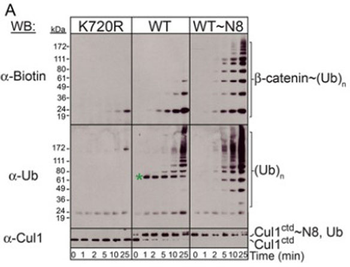
Western Blot of Anti-Biotin antibody. Conformational control of CRL activities. (A) Polyubiquitination reactions with SCFβTRCP/β-catenin phosphopeptide (left), and SCFSkp2/CksHs1/ phospho-p27 (right) reconstituted with non-NEDD8ylatable (K720R), un-NEDD8ylated wild-type (WT), and fully NEDD8ylated Cul1-Rbx1 (WT~N8). Reaction products were detected by immunoblotting, top panels with anti-biotin (left) or anti-p27 (right), middle with anti-His (Ubiquitin; green * - Cul1~Ubiquitin), and lower with anti-Cul1 C-terminus antisera.

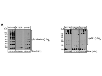
Western Blot of Anti-Biotin Antibody. Functional analysis of mutations influencing RING conformational flexibility. (A) Time-courses of polyubiquitination by SCFs reconstituted with fully-NEDD8ylated wild-type Cul1-Rbx1 (WT~N8), the non-NEDD8ylatable control (K720R), and the Cul1-Rbx1 WHB deletion mutant (ΔWHB). Left - SCFβTRCP-mediated polyubiquitination of a biotin-labeled β-catenin phosphopeptide, detected by western blotting with anti-biotin antisera. Right - SCFSkp2/CksHs1-mediated polyubiquitination of phospho-p27, detected by western blotting with anti-p27 antisera.
Documents Download
Request a Document
Protocol Information
Biotin Antibody (orb750609)
Participating in our Biorbyt product reviews program enables you to support fellow scientists by sharing your firsthand experience with our products.
Login to Submit a Review