You have no items in your shopping cart.
Description
Research Area
Cell Biology
Images & Validation
−Item 1 of 4
| Tested Applications | IF, IHC-P, WB |
|---|---|
| Dilution Range | IF - 1:25, WB - 1:500-2000, IHC-P - 1:25 |
| Reactivity | Human |
Key Properties
−| Host | Mouse |
|---|---|
| Clonality | Monoclonal |
| Isotype | IgG1,k |
| Clone No. | B3731EV861X30X69 |
| Immunogen | This BID antibody is generated from a mouse immunized with a recombinant protein of human BID. |
| Target | BID |
| Molecular Weight | 21995 Da |
| Conjugation | Unconjugated |
Storage & Handling
−| Storage | Maintain refrigerated at 2-8°C for up to 2 weeks. For long term storage store at -20°C in small aliquots to prevent freeze-thaw cycles |
|---|---|
| Form/Appearance | Purified monoclonal antibody supplied in PBS with 0.09% (W/V) sodium azide. This antibody is purified through a protein G column, followed by dialysis against PBS. |
| Expiration Date | 12 months from date of receipt. |
| Disclaimer | For research use only |
Alternative Names
−BH3-interacting domain death agonist, p22 BID, BID, BH3-interacting domain death agonist p15, p15 BID, BH3-interacting domain death agonist p13, p13 BID, BH3-interacting domain death agonist p11, p11 BID, BID
Similar Products
−- Item 1 of 7
Bid Antibody (BH3 Domain Specific) [orb1936953]
IHC-P, WB
Human, Mouse
Rabbit
Polyclonal
Unconjugated
50 μl, 100 μl - Item 1 of 7
BID Rabbit Polyclonal Antibody [orb182359]
FC, ICC, IF, IHC, WB
Human, Mouse, Rat
Rabbit
Polyclonal
Unconjugated
100 μg - Item 1 of 5
Bid Rabbit Polyclonal Antibody [orb10188]
FC, ICC, IF, IHC-Fr, IHC-P
Mouse, Rat
Human, Mouse, Rat
Rabbit
Polyclonal
Unconjugated
50 μl, 100 μl, 200 μl - Item 1 of 5
- Item 1 of 1
Human BH3 Interacting Domain Death Agonist (Bid) ELISA Kit [orb775567]
Human
1.57-100 ng/mL
0.56 ng/mL
96 T, 48 T


Quality Guarantee
Explore bioreagents carefree to elevate your research. All our products are rigorously tested for performance. If a product does not perform as described on its datasheet, our scientific support team will provide expert troubleshooting, a prompt replacement, or a refund. For full details, please see our Terms & Conditions and Buying Guide. Contact us at [email protected].

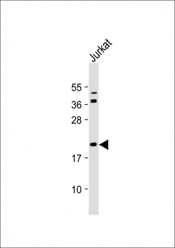
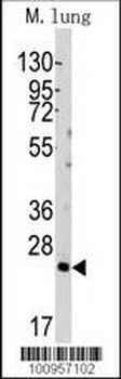
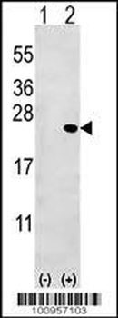
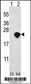
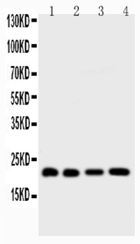
All lanes: Anti-BID Antibody at 1:4000 dilution. Lane 1: Jurkat whole cell lysates. Lane 2: A431 whole cell lysates. Lane 3: THP-1 whole cell lysates. Lane 4: 293T/17 whole cell lysates. Lysates/proteins at 20 μg per lane. Secondary Goat Anti-mouse IgG, (H+L), Peroxidase conjugated at 1/10000 dilution. Predicted band size: 22 kDa. Blocking/Dilution buffer: 5% NFDM/TBST.
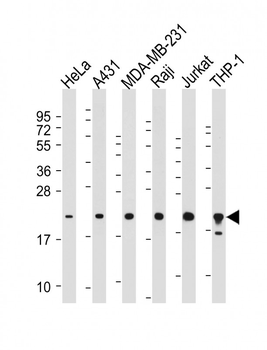
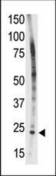
All lanes: Anti-BID Antibody at 1:500-2000 dilution. Lane 1: HeLa whole cell lysate. Lane 2: A431 whole cell lysate. Lane 3: MDA-MB-231 whole cell lysate. Lane 4: Raji whole cell lysate. Lane 5: Jurkat whole cell lysate. Lane 6: THP-1 whole cell lysate. Lysates/proteins at 20 µg per lane. Secondary Goat Anti-mouse IgG, (H+L), Peroxidase conjugated at 1/10000 dilution. Predicted band size: 22 kDa. Blocking/Dilution buffer: 5% NFDM/TBST.
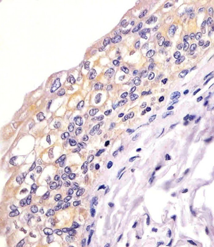
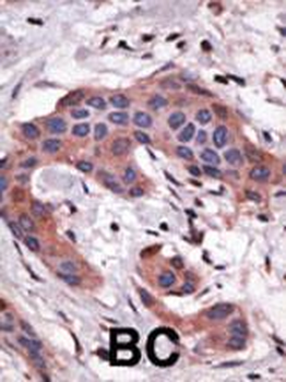
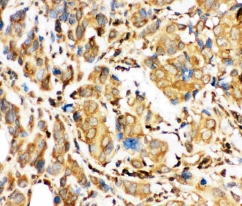
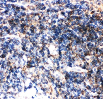
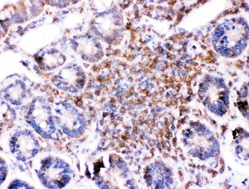
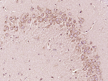
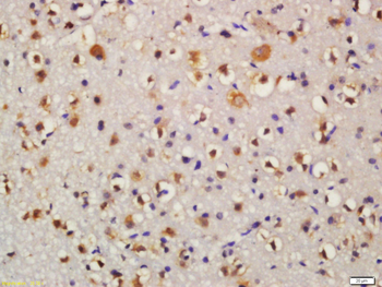
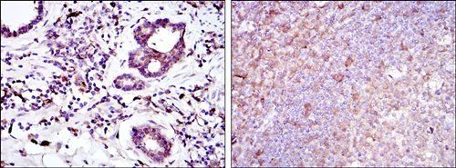
Staining BID in human bladder sections by Immunohistochemistry (IHC-P - paraformaldehyde-fixed, paraffin-embedded sections). Tissue was fixed with formaldehyde and blocked with 3% BSA for 0.5 hour at room temperature; antigen retrieval was by heat mediation with a citrate buffer (pH6). Samples were incubated with primary antibody (1/25) for 1 hours at 37°C. A undiluted biotinylated goat polyvalent antibody was used as the secondary antibody.
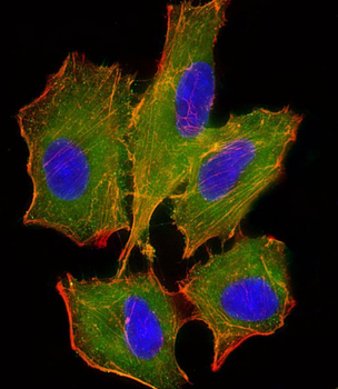
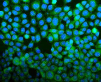
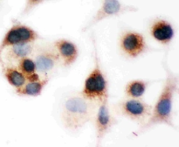
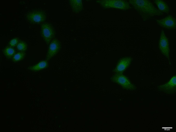
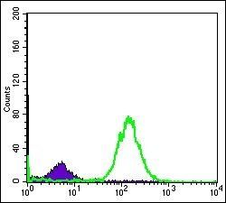
Immunofluorescent analysis of 4% paraformaldehyde-fixed, 0.1% Triton X-100 permeabilized A549 (human lung adenocarcinoma epithelial cell line) cells labeling BID at 1/25 dilution, followed by Dylight 488-conjugated goat anti-mouse IgG secondary antibody at 1/200 dilution (green). Immunofluorescence image showing cytoplasm staining on A549 cell line. Cytoplasmic actin is detected with Dylight 554 Phalloidin at 1/100 dilution (red).The nuclear counter stain is DAPI (blue).
Quick Database Links
Gene Symbol
BID
UniProt
UniProt Details
− No UniProt data available
Documents Download
Datasheet
Product Information
Request a Document
Protocol Information
WB
Western Blot (IB, immunoblot)
IHC-P
Immunohistochemistry Paraffin
IF
Immunofluorescence
BID Antibody (orb1926251)

- 0.0
Based on 0 reviews
Participating in our Biorbyt product reviews program enables you to support fellow scientists by sharing your firsthand experience with our products.
Login to Submit a ReviewAvailable Sizes
Select a size below
Choose Conjugation or Carrier Free Version
Free Secondary Antibody (20 ul)0/0
Please add an antibody product to your cart first.