You have no items in your shopping cart.
Description
Research Area
Cancer Biology; Musculoskeletal & Connective Tissue Research
Images & Validation
−Item 1 of 6
| Tested Applications | IHC-P, WB |
|---|---|
| Dilution Range | WB - 1:4000-1:8000, IHC - 1:50, IHC-P - 1:25 |
| Reactivity | Human, Mouse, Rat |
Key Properties
−| Host | Mouse |
|---|---|
| Clonality | Monoclonal |
| Isotype | IgG |
| Clone No. | B0J32F32 |
| Immunogen | ACTB recombinant protein is used to produce this monoclonal antibody. Antigen Region: Unknown. |
| Target | ACTB |
| Molecular Weight | 41737 Da |
| Conjugation | Unconjugated |
Storage & Handling
−| Storage | Maintain refrigerated at 2-8°C for up to 2 weeks. For long term storage store at -20°C in small aliquots to prevent freeze-thaw cycles |
|---|---|
| Form/Appearance | Purified monoclonal antibody supplied in PBS with 0.09% (W/V) sodium azide. This antibody is purified through a protein G column, followed by dialysis against PBS. |
| Expiration Date | 12 months from date of receipt. |
| Disclaimer | For research use only |
Alternative Names
−Actin, cytoplasmic 1, Beta-actin, Actin, cytoplasmic 1, N-terminally processed, ACTB
Similar Products
−- Item 1 of 5
beta Actin Rabbit Polyclonal Antibody [orb10033]
ELISA, IHC-P, WB
Human, Mouse, Rat
Rabbit
Polyclonal
Unconjugated
100 μg, 20 μg - Item 1 of 7
beta Actin Rabbit Polyclonal Antibody [orb181785]
ELISA, ICC, IF, IHC-P, WB
Human, Mouse
Rabbit
Polyclonal
Unconjugated
100 μg - Item 1 of 5
Beta-Actin Rabbit Polyclonal Antibody (Loading Control) [orb500817]
FC, ICC, IF, IHC-Fr, IHC-P, WB
Canine, Feline, Fish, Gallus, Guinea pig, Hamster, Insect, Porcine, Rabbit, Sheep
Human, Mouse, Rat
Rabbit
Polyclonal
Unconjugated
100 μl, 500 μl, 200 μl, 1 ml - Item 1 of 10
ACTB Antibody [orb420309]
ELISA, IF, KO/KD Validated, WB
Frog, Human, Mouse
Rabbit
Polyclonal
Unconjugated
200 μg - Item 1 of 10
ACTB Antibody [orb420310]
ELISA, IF, KO/KD Validated, WB
Frog, Human, Mouse
Rabbit
Polyclonal
Unconjugated
25 μl


Quality Guarantee
Explore bioreagents carefree to elevate your research. All our products are rigorously tested for performance. If a product does not perform as described on its datasheet, our scientific support team will provide expert troubleshooting, a prompt replacement, or a refund. For full details, please see our Terms & Conditions and Buying Guide. Contact us at [email protected].
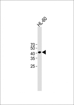
All lanes: Anti-Beta-actin Antibody at 1:2000 dilution + HL-60 whole cell lysate. Lysates/proteins at 20 µg per lane. Secondary: Goat Anti-Mouse IgG, (H+L), Peroxidase conjugated at 1/8000 dilution.Observed band size: 42 KDa. Blocking/Dilution buffer: 5% NFDM/TBST.
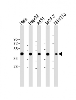
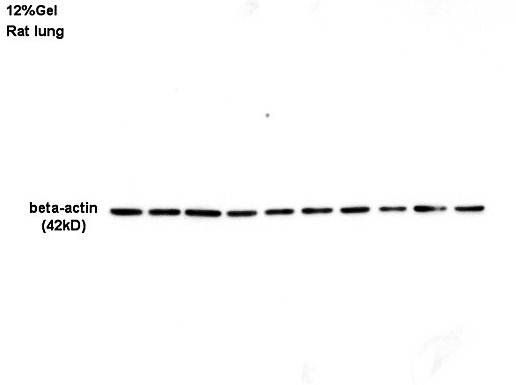
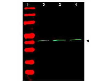
All lanes: Anti-Beta-actin Antibody at 1:4000-1:8000 dilution. Lane 1: Hela whole cell lysate. Lane 2: HepG2 whole cell lysate. Lane 3: A431 whole cell lysate. Lane 4: MCF-7 whole cell lysate. Lane 5: NIH/3T3 whole cell lysate.Lysates/proteins at 20 µg per lane. Secondary Goat Anti-mouse IgG, (H+L), Peroxidase conjugated at 1/10000 dilution. Predicted band size: 42 kDa. Blocking/Dilution buffer: 5% NFDM/TBST.
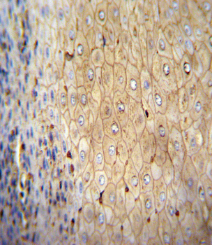
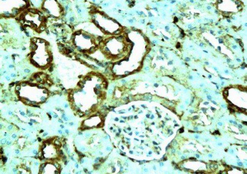
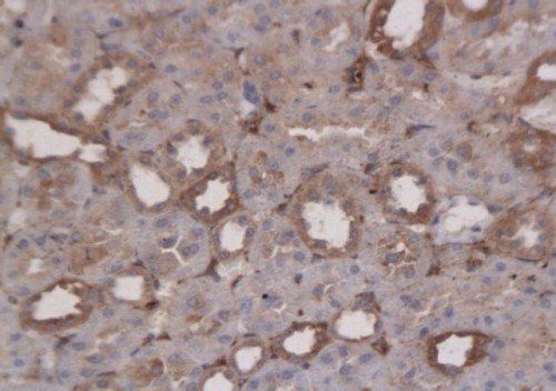
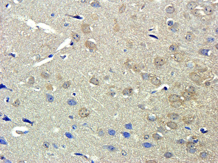
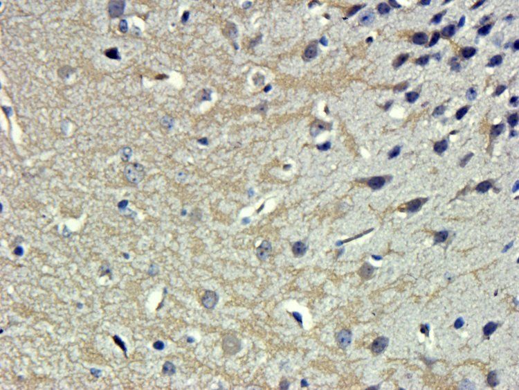
Immunohistochemical analysis of paraffin-embedded H. spleen section using Beta-actin Antibody. diluted at 1:25 dilution. A peroxidase-conjugated goat anti-Mouse IgG at 1:400 dilution was used as the secondary antibody, followed by DAB staining.
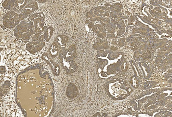
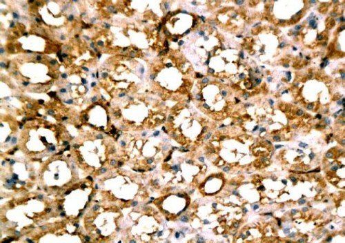
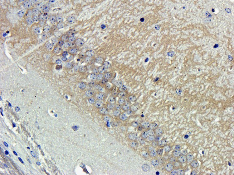
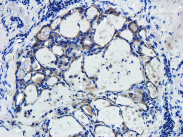
Immunohistochemical analysis of paraffin-embedded Human Ovarian cancer section using Pink1. diluted at 1:50 dilution. A undiluted biotinylated goat polyvalent antibody was used as the secondary, followed by DAB staining.
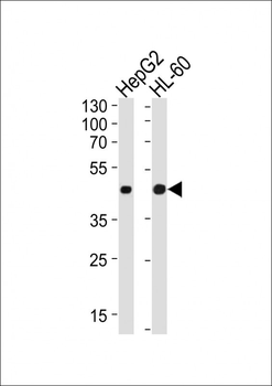
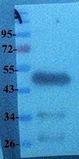
Western blot analysis of lysates from HepG2, HL-60 cell line (from left to right), using Beta-actin Antibody. diluted at 1:1000 at each lane. A goat anti-mouse IgG H&L (HRP) at 1:10000 dilution was used as the secondary Antibody. Lysates at 20 μg per lane.
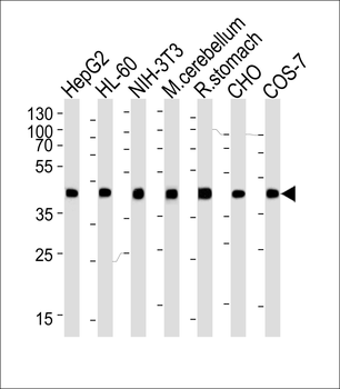
Western blot analysis of lysates from HepG2, HL-60, mouse NIH/3T3 cell line, mouse cerebellum and rat stomach tissue lysate, CHO, COS-7 cell line lysate (from left to right), using Beta-actin Antibody. diluted at 1:1000 at each lane. A goat anti-mouse IgG H&L (HRP) at 1:3000 dilution was used as the secondary Antibody. Lysates at 35 μg per lane.
Quick Database Links
UniProt Details
− No UniProt data available
NCBI Reference Sequences
−Associated Accession Numbers
Curated reference sequences for the gene transcript and protein product| Protein | NP_001092.1 |
|---|
Documents Download
Datasheet
Product Information
Request a Document
Protocol Information
WB
Western Blot (IB, immunoblot)
IHC-P
Immunohistochemistry Paraffin
Beta-actin Antibody (orb1939450)

- 0.0
Based on 0 reviews
Participating in our Biorbyt product reviews program enables you to support fellow scientists by sharing your firsthand experience with our products.
Login to Submit a ReviewAvailable Sizes
Select a size below
Choose Conjugation or Carrier Free Version
Free Secondary Antibody (20 ul)0/0
Please add an antibody product to your cart first.