You have no items in your shopping cart.
Featured
KO/KD
Validated
Validated
Description
Research Area
Cell Biology
Images & Validation
−Item 1 of 10
| Tested Applications | ELISA, ICC, IF, IHC-P, KO/KD Validated, WB |
|---|---|
| Reactivity | Human, Mouse |
| Predicted Reactivity | Bovine, Guinea pig, Rat |
| Application Notes |
Key Properties
−| Antibody Type | Primary Antibody |
|---|---|
| Host | Rabbit |
| Clonality | Polyclonal |
| Isotype | IgG |
| Immunogen | Anti-BACE antibody (orb1239248) was raised against a peptide corresponding to 17 amino acids near the carboxy terminus of human BACE. The immunogen is located within the last 50 amino acids of BACE. |
| Target | BACE1 |
| Molecular Weight | Predicted: 55kDObserved: 65kD (Post-modification: 4 N-linked glycosylation) |
| Purification | BACE Antibody is affinity chromatography purified via peptide column. |
| Conjugation | Unconjugated |
Storage & Handling
−| Storage | Maintain refrigerated at 2-8°C for up to 2 weeks. For long term storage store at -20°C in small aliquots to prevent freeze-thaw cycles. |
|---|---|
| Form/Appearance | Liquid |
| Buffer/Preservatives | BACE Antibody is supplied in PBS containing 0.02% sodium azide. |
| Concentration | 1 mg/mL |
| Expiration Date | 12 months from date of receipt. |
| Disclaimer | For research use only |
Alternative Names
−BACE Antibody: ASP2, BACE, HSPC104, KIAA1149, Beta-secretase 1, Aspartyl protease 2, ASP2
Similar Products
−- Item 1 of 4
BACE1 Rabbit Polyclonal Antibody [orb10164]
ICC, IF, IHC-Fr, IHC-P, WB
Canine, Gallus, Guinea pig, Porcine, Rabbit
Human, Mouse, Rat
Rabbit
Polyclonal
Unconjugated
50 μl, 100 μl, 200 μl - Item 1 of 6
BACE1 Rabbit Polyclonal Antibody [orb579899]
IF, IHC, WB
Bovine, Canine, Equine, Guinea pig, Rabbit, Rat, Zebrafish
Human, Mouse
Rabbit
Polyclonal
Unconjugated
100 μl - Item 1 of 1
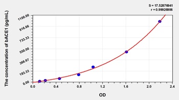
Rat Beta-Site APP Cleaving Enzyme 1 (bACE1) ELISA Kit [orb777177]
Rat
15.63-1000 pg/mL
6.7 pg/mL
48 T, 96 T - Item 1 of 1
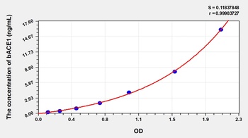
Human Beta-Site APP Cleaving Enzyme 1 (bACE1) ELISA Kit [orb775893]
Human
0.25-16 ng/mL
0.11 ng/mL
48 T, 96 T - Item 1 of 1
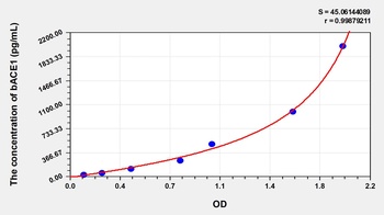
Mouse Beta-Site APP Cleaving Enzyme 1 (bACE1) ELISA Kit [orb777389]
Mouse
31.25-2000 pg/mL
14.3 pg/mL
48 T, 96 T

Quality Guarantee
Explore bioreagents carefree to elevate your research. All our products are rigorously tested for performance. If a product does not perform as described on its datasheet, our scientific support team will provide expert troubleshooting, a prompt replacement, or a refund. For full details, please see our Terms & Conditions and Buying Guide. Contact us at [email protected].
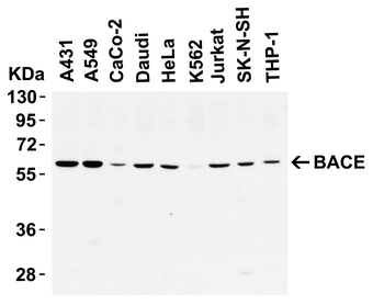
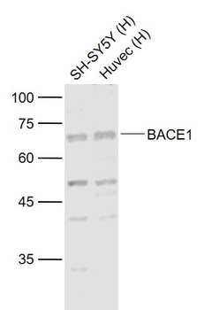
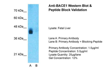
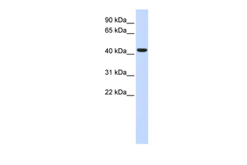
WB Validation in Human Cell Lines. Loading: 10 μg of lysate. Antibodies: BACE, orb1239248, 1 μg/mL, 1 h incubation at RT in 5% NFDM/TBST. Secondary: Goat Anti-Rabbit IgG HRP conjugate at 1:10000 dilution.
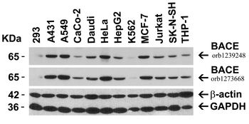
Independent Antibody Validation (IAV) via Protein Expression Profile in Cell Lines. Loading: 15 μg of lysates per lane. Antibodies: BACE orb1239248 (1 μg/mL), BACE orb1273668 (1 μg/mL), beta-actin (1 μg/mL), and GAPDH (0.02 μg/mL), 1h incubation at RT in 5% NFDM/TBST. Secondary: Goat anti-rabbit IgG HRP conjugate at 1:10000 dilution.
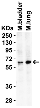
WB Validation in Mouse Tissues. Loading: 15 μg of lysate. Antibodies: BACE, orb1239248, 2 μg/mL, 1 h incubation at RT in 5% NFDM/TBST. Secondary: Goat Anti-Rabbit IgG HRP conjugate at 1:10000 dilution.
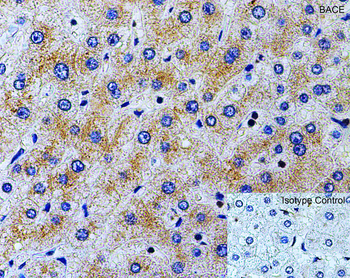
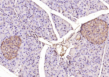
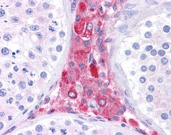
Immunohistochemistry Validation of BACE in Human Liver. Immunohistochemical analysis of paraffin-embedded human liver tissue using anti-BACE antibody (orb1239248) at 2 μg/ml. Tissue was fixed with formaldehyde and blocked with 10% serum for 1 h at RT; antigen retrieval was by heat mediation with a citrate buffer (pH6). Samples were incubated with primary antibody overnight at 4°C. A goat anti-rabbit IgG H&L (HRP) at 1/250 was used as secondary. Counter stained with Hematoxylin.
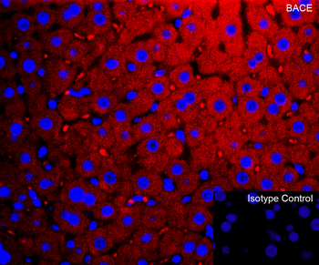
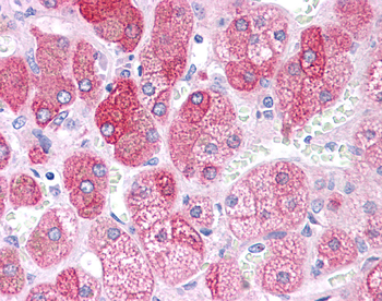
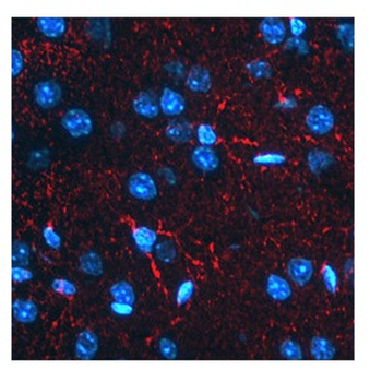
Immunofluorescence Validation of BACE in Mouse Liver. Immunofluorescent analysis of 4% paraformaldehyde-fixed mouse liver tissue labeling BACE with orb1239248 at 10 μg/mL, followed by goat anti-rabbit IgG secondary antibody at 1/500 dilution (red) and DAPI staining (blue).
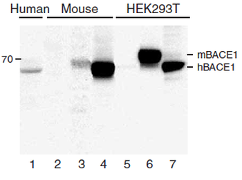
KO and Overexpression Validation of BACE in Human and Mouse Brain and 293 Cells. Western blot analysis of the BACE1 (orb1239248) antibody's ability to recognize human and murine BACE1. The BACE1 antibody recognized both the mouse and human forms of BACE1. Lanes 1–4 are frontal cortex homogenates from human and mouse brains. Lane 1 is from a neurologically unimpaired aged human control case, lane 2 from a BACE1-deficient mouse, lane 3 from a nontransgenic mouse and lane 4 from hBACE1 transgenic mouse. Lanes 5–7 are lysates from HEK293T cells transfected with a plasmid vector expressing eGFP, mBACE1 and hBACE1, respectively.
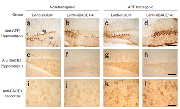
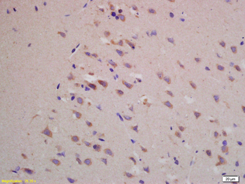
KD Validation of BACE in Mouse Brain. Characterization of the effects of lenti-siBACE1-6 expression in the brains of APP transgenic mice. (a–d) Anti-eGFP immunoreactivity in the hippocampus (the injection site) shows comparable and consistent expression of lenti-siRNA constructs in the dentate gyrus (dg) and stratus polymorphus (sp). (e) Anti-BACE1 immunoreactivity in the hippocampus of nontransgenic mice treated with lenti-siGlut4. (f) Reduced BACE1 immunostaining in the hippocampus of nontransgenic mice treated with lenti-siBACE1-6 vector. (g) Intense BACE1 immunoreactivity in the hippocampus of APP transgenic mice treated with lenti-siGlut4. (h) Reduced BACE1 expression in APP transgenic mice treated with lenti-siBACE1-6 vector. (i, j) Anti-BACE1 reacted with pyramidal cell bodies in the neocortex, which was not injected.
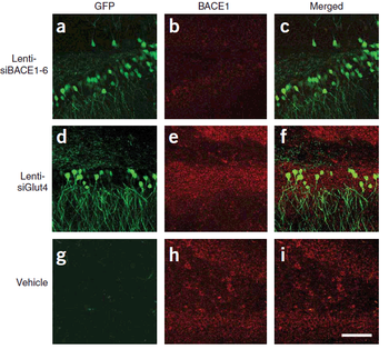
KD Validation of BACE in Mouse Brain. Immunolabeling patterns of BACE1 expression and the lenti-siRNA distribution. Sections from APP transgenic mice treated with the eGFPtagged lenti siRNA vectors (green) were co-immunolabeled with an antibody against BACE1 (red) and imaged with the LSCM. All sections are from the hippocampus of treated mice. (a–c) Lenti-siBACE1-6–treated mice. Areas within the hippocampus expressing the eGFP tagged vector have reduced BACE1 immunolabeling. (d–f) Mice treated with the eGFP-tagged control lenti-siGlut4 show unchanged expression of BACE1 in the hippocampus. (g–i) Mice treated with a saline vehicle show unchanged expression of BACE1 in the hippocampus.
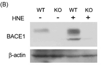
KO Validation of BACE in MEF Cells. Wildtype and BACE -/- MEFs were exposed to HNE (15_M) for 2 h. BACE1 levels were examined by Western blot with anti-BACE antibodies (orb1239248).
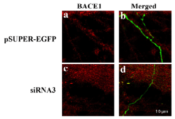
KD Validation of BACE in DRG (Hyun, orb1239261). Decreased BACE1 expression in DRG following siRNA3 transfection. DRG neurons were transfected with 1 μg siRNA3 plasmid and incubated for 48 hours in 37°C. DRG neurons were stained for BACE1 using the Anti-BACE antibody. (a, b) Neurons transfected with the control plasmid pSUPER-EGFP (green) did not display any changes in BACE1 expression (red). (c, d) DRG neurons transfected with siR¬NA3 displayed reduced BACE1 expression in the axon.
Documents Download
Datasheet
Product Information
Request a Document
Protocol Information
WB
Western Blot (IB, immunoblot)
IHC-P
Immunohistochemistry Paraffin
IF
Immunofluorescence
ICC
Immunocytochemistry
ELISA
Enzyme-linked Immunosorbent Assay (EIA)
BACE1 Antibody (orb1239248)

- 0.0
Based on 0 reviews
Participating in our Biorbyt product reviews program enables you to support fellow scientists by sharing your firsthand experience with our products.
Login to Submit a ReviewAvailable Sizes
Select a size below
Free Secondary Antibody (20 ul)0/0
Please add an antibody product to your cart first.