You have no items in your shopping cart.
Description
Research Area
Infectious Disease & Virology
Images & Validation
−Item 1 of 4
| Tested Applications | ELISA, FC, ICC, IHC-P, IP, WB |
|---|---|
| Reactivity | Human, Primate |
| Application Notes |
Key Properties
−| Antibody Type | Primary Antibody |
|---|---|
| Clonality | Monoclonal |
| Isotype | Mouse IgG1 kappa |
| Clone No. | 4S.B3 |
| Immunogen | Interferon gamma derived from human leukocytes |
| Target | IFN gamma |
| Purification | Purified antibody is conjugated with biotin LC-NHS ester under optimum conditions and unconjugated antibody and free biotin are removed by size-exclusion chromatography. |
| Conjugation | Biotin |
Storage & Handling
−| Storage | Store at 2-8°C. Do not freeze. |
|---|---|
| Buffer/Preservatives | Stabilizing phosphate buffered saline (PBS), pH 7.4, 15 mM sodium azide |
| Concentration | 1 mg/ml |
| Expiration Date | 12 months from date of receipt. |
| Disclaimer | For research use only |
Alternative Names
−Interferon gamma, IFN-gamma
Similar Products
−- Item 1 of 1
- Item 1 of 1
- Item 1 of 4


Quality Guarantee
Explore bioreagents carefree to elevate your research. All our products are rigorously tested for performance. If a product does not perform as described on its datasheet, our scientific support team will provide expert troubleshooting, a prompt replacement, or a refund. For full details, please see our Terms & Conditions and Buying Guide. Contact us at [email protected].
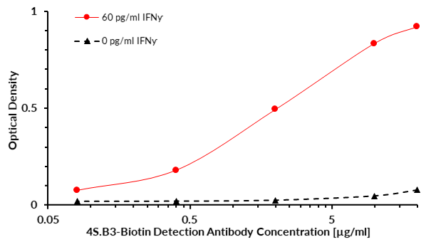
Cytokine IFN gamma (IFNƴ) sandwich ELISA (enzyme-linked immunosorbent assay) that utilizes antibody clone NIB42 as the protein capture antibody and antibody clone 4S.B3-Biotin as the detection antibody. Recombinant IFNƴ was captured by NIB42 and detected by variable amounts of 4S.B3-Biotin. The 4S.B3-Biotin conjugate was visualized by Streptavidin-HRP and substrate solution (TMB). The intensity of the signal is proportional to the amount of cytokine and to the amount of the detection antibody.
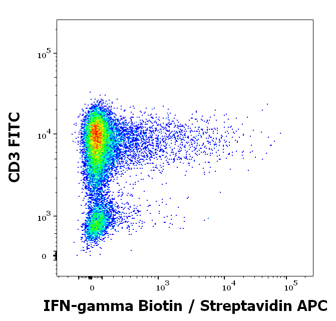
Flow cytometry multicolor intracellular staining pattern of human lymphocytes (PHA stimulated and Brefeldin A + Monesin treated) stained using anti-human IFN-gamma (4S.B3) biotin antibody (concentration in sample 1.7 μg/ml, Streptavidin APC) and anti-human CD3 (UCHT1) FITC antibody (20 μl reagent / 100 μl of peripheral whole blood).
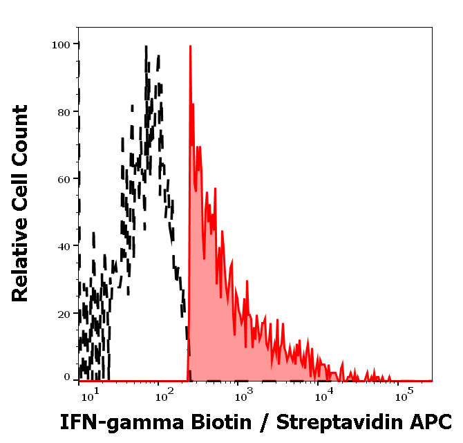
Separation of human IFN-gamma positive CD3 positive lymphocytes (red-filled) from IFN-gamma negative CD3 negative lymphocytes (black-dashed) in flow cytometry analysis (intracellular staining) of human peripheral whole blood (PHA stimulated and Brefeldin A + Monesin treated) stained using anti-human IFN-gamma (4S.B3) biotin antibody (concentration in sample 1.7 μg/ml, Streptavidin APC).
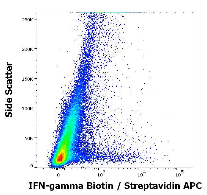
Flow cytometry intracellular staining pattern of human peripheral whole blood (PHA stimulated and Brefeldin A + Monesin treated) stained using anti-human IFN-gamma (4S.B3) biotin antibody (concentration in sample 1.7 μg/ml, Streptavidin APC).
Documents Download
Datasheet
Product Information
Request a Document
Protocol Information
WB
Western Blot (IB, immunoblot)
IHC-P
Immunohistochemistry Paraffin
FC
Flow Cytometry
ICC
Immunocytochemistry
ELISA
Enzyme-linked Immunosorbent Assay (EIA)
IP
Immunoprecipitation
IFN gamma Antibody (Biotin) (orb179917)

- 0.0
Based on 0 reviews
Participating in our Biorbyt product reviews program enables you to support fellow scientists by sharing your firsthand experience with our products.
Login to Submit a ReviewAvailable Sizes
Select a size below
Free Secondary Antibody (20 ul)0/0
Please add an antibody product to your cart first.