You have no items in your shopping cart.
Description
Research Area
Immunology & Inflammation
Images & Validation
−Item 1 of 4
| Tested Applications | FC, IP, WB |
|---|---|
| Reactivity | Human |
| Application Notes |
Key Properties
−| Antibody Type | Primary Antibody |
|---|---|
| Clonality | Monoclonal |
| Isotype | Mouse IgG1 |
| Clone No. | MEM-240 |
| Immunogen | Recombinant Vaccinia virus encoding CD222. |
| Target | CD222 |
| Purification | Purified by protein-A affinity chromatography. |
| Conjugation | Unconjugated |
Storage & Handling
−| Storage | Maintain refrigerated at 2-8°C for up to 2 weeks. For long term storage store at -20°C in small aliquots to prevent freeze-thaw cycles. |
|---|---|
| Buffer/Preservatives | Phosphate buffered saline (PBS), pH 7.4, 15 mM sodium azide |
| Concentration | 1 mg/ml |
| Expiration Date | 12 months from date of receipt. |
| Disclaimer | For research use only |
Alternative Names
−IGF2R, MPR1, CIMPR, MPR300, M6P-R
Similar Products
−- Item 1 of 7
CD222 Rabbit Polyclonal Antibody [orb385612]
ICC, IF, IHC-P, WB
Guinea pig, Human, Mouse, Rat
Rabbit
Polyclonal
Unconjugated
100 μg - Item 1 of 7
Mannose 6 Phosphate Receptor/IGF2R Rabbit Polyclonal Antibody [orb413019]
ELISA, FC, ICC, IF, IHC, WB
Human, Mouse, Rat
Rabbit
Polyclonal
Unconjugated
100 μg - Item 1 of 6
- Item 1 of 1
Human Insulin Like Growth Factor 2 Receptor (IGF2R) ELISA Kit [orb775992]
Human
0.63-40 ng/mL
0.224 ng/mL
96 T, 48 T - Item 1 of 5
IGF2R Mouse Monoclonal Antibody [orb763094]
FC, ICC, IF, IHC, WB
Human
Mouse
Monoclonal
Unconjugated
100 μg

Quality Guarantee
Explore bioreagents carefree to elevate your research. All our products are rigorously tested for performance. If a product does not perform as described on its datasheet, our scientific support team will provide expert troubleshooting, a prompt replacement, or a refund. For full details, please see our Terms & Conditions and Buying Guide. Contact us at [email protected].
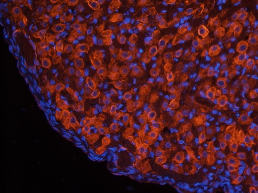
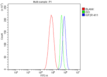
Flow cytometry detection of CD222 in human peripheral blood with anti-CD222 (MEM-240) purified, GAM-APC.
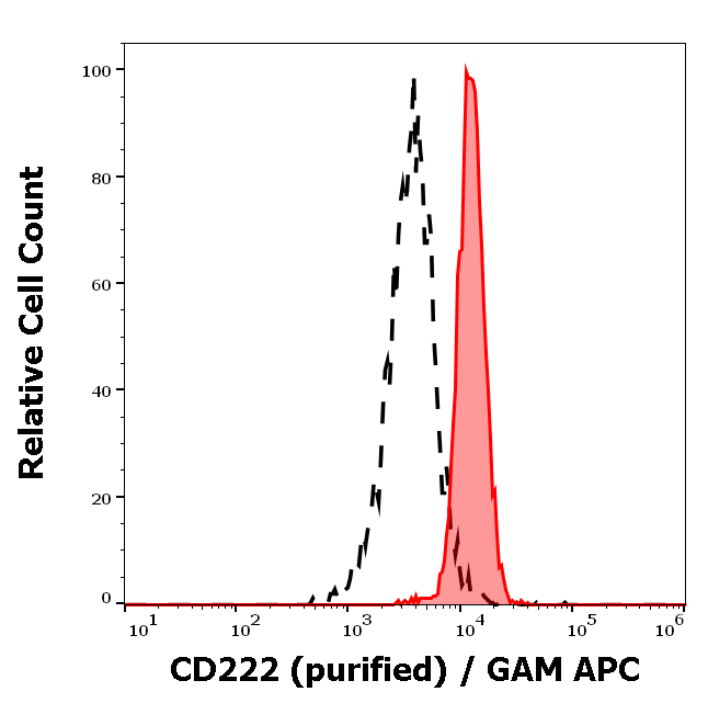
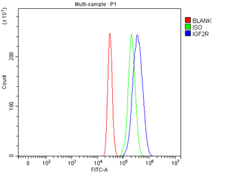
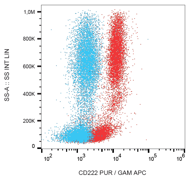
Separation of human neutrophil granulocytes (red-filled) from lymphocytes (black-dashed) in flow cytometry analysis (surface staining) of human peripheral whole blood stained using anti-human CD222 (MEM-240) purified antibody (concentration in sample 3 µg/ml) GAM APC.
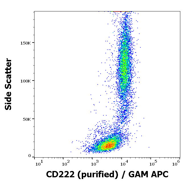
Flow cytometry surface staining pattern of human peripheral whole blood stained using anti-human CD222 (MEM-240) purified antibody (concentration in sample 3 µg/ml) GAM APC.
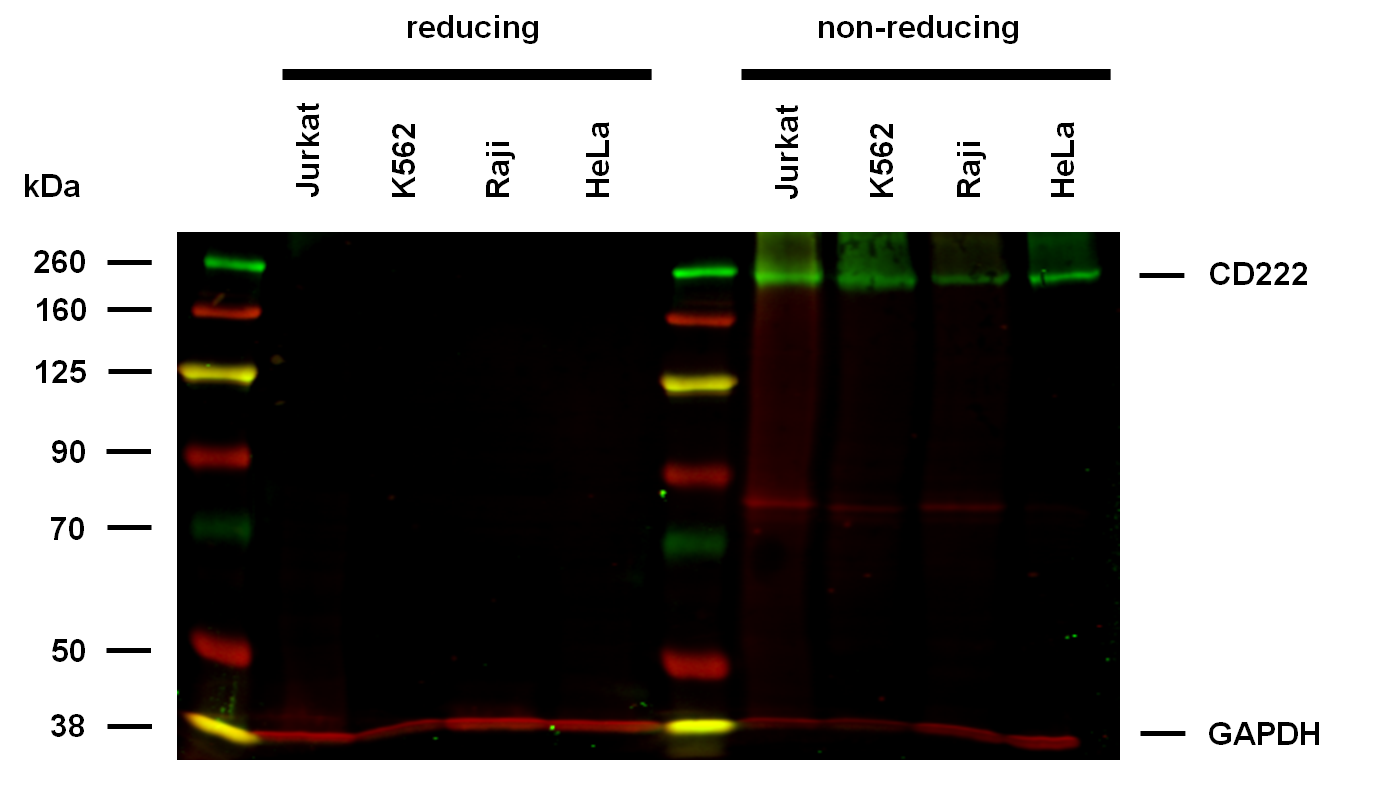
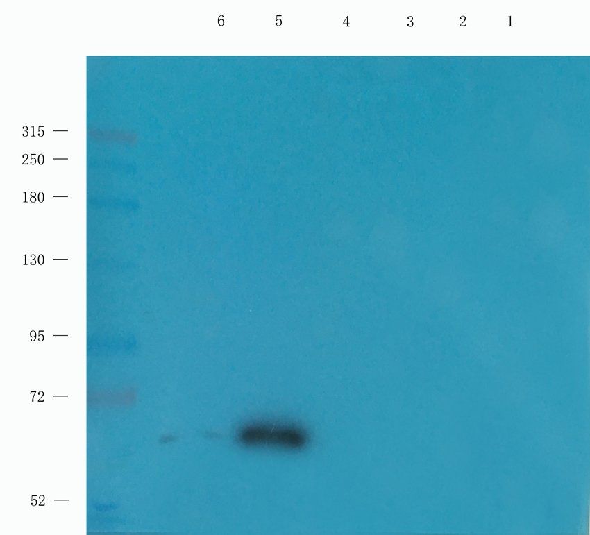
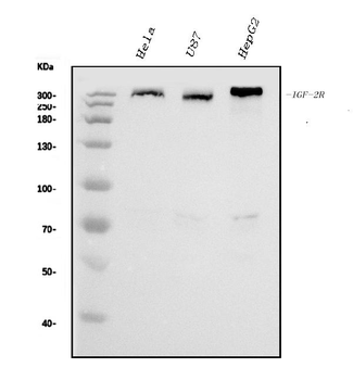
Anti-Hu CD222 Purified (clone MEM-240) works in WB application under non-reducing conditions. Western blotting analysis was performed on whole cell extracts (RIPA lysis buffer) of Jurkat, K562, Raji, and HeLa cell lines, mixed and heated (100°C, 5 min) with reducing and non-reducing SDS-loading buffer. Samples were resolved using 7% Tris-glycine SDS gel electrophoresis. Nitrocellulose membrane blot was probed with mouse IgG1 monoclonal antibody MEM-240 (1 µg/ml), followed by IRDye 800CW Goat-anti-Mouse IgG (green). Mouse anti-GAPDH monoclonal antibody FF26A conjugated with DyLight 680 (0.1 µg/ml), was used as the loading control (red). Multiplex fluorescent Western blot detection was performed. CD222 molecules were detected at ~250 kDa in all analysed cell lines.
Documents Download
Datasheet
Product Information
Request a Document
Protocol Information
WB
Western Blot (IB, immunoblot)
FC
Flow Cytometry
IP
Immunoprecipitation
CD222 Antibody (orb402817)

- 0.0
Based on 0 reviews
Participating in our Biorbyt product reviews program enables you to support fellow scientists by sharing your firsthand experience with our products.
Login to Submit a ReviewAvailable Sizes
Select a size below
Free Secondary Antibody (20 ul)0/0
Please add an antibody product to your cart first.