You have no items in your shopping cart.
Description
Research Area
Epigenetics & Chromatin
Images & Validation
−Item 1 of 5
| Tested Applications | ELISA, FC, ICC, IP, WB |
|---|---|
| Reactivity | Human |
| Application Notes |
Key Properties
−| Antibody Type | Primary Antibody |
|---|---|
| Clonality | Monoclonal |
| Isotype | Mouse IgG2b kappa |
| Clone No. | 236 |
| Immunogen | human HDAC6 |
| Target | HDAC6 |
| Purification | Purified by protein-A affinity chromatography. |
| Conjugation | Unconjugated |
Storage & Handling
−| Storage | Maintain refrigerated at 2-8°C for up to 2 weeks. For long term storage store at -20°C in small aliquots to prevent freeze-thaw cycles. |
|---|---|
| Buffer/Preservatives | Phosphate buffered saline (PBS), pH 7.4, 15 mM sodium azide |
| Concentration | 1 mg/ml |
| Expiration Date | 12 months from date of receipt. |
| Disclaimer | For research use only |
Alternative Names
−Histone deacetylase 6
Similar Products
−- Item 1 of 6
Hdac6 Rabbit Polyclonal Antibody [orb574130]
IHC, WB
Guinea pig, Rat
Human, Mouse
Rabbit
Polyclonal
Unconjugated
100 μl - Item 1 of 4
HDAC6 Antibody [orb154549]
ELISA, FC, IF, IHC
Bovine, Canine, Mouse, Rat
Human
Polyclonal
Unconjugated
100 μg - Item 1 of 4
HDAC6 rabbit pAb Antibody [orb765379]
ELISA, IF, IHC, WB
Human, Mouse
Polyclonal
Unconjugated
50 μl, 100 μl - Item 1 of 4
HDAC6 Antibody [orb1410250]
FC, IF
Human
Mouse
Monoclonal
Unconjugated
20 μg, 100 μg, 100 μg (without BSA and Azide) - Item 1 of 4
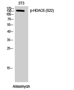
HDAC6 (phospho Ser22) rabbit pAb Antibody [orb767279]
ELISA, IF, IHC, WB
Human, Mouse
Polyclonal
Unconjugated
50 μl, 100 μl


Quality Guarantee
Explore bioreagents carefree to elevate your research. All our products are rigorously tested for performance. If a product does not perform as described on its datasheet, our scientific support team will provide expert troubleshooting, a prompt replacement, or a refund. For full details, please see our Terms & Conditions and Buying Guide. Contact us at [email protected].
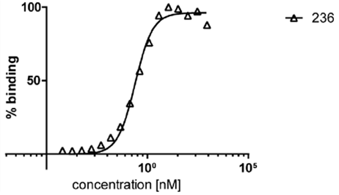
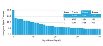
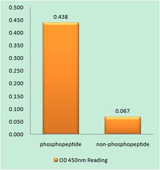
ELISA analysis of human HDAC6 by mouse monoclonal antibody 236. MaxiSorp plate was coated with recombinant human HDAC6, then blocked with BSA, and exposed to dilution series of anti-HDAC6 primary antibody, followed by HRP-conjugated anti-mouse secondary antibody, and colorimetric signal of processed OPD substrate was measured at 492 nm.
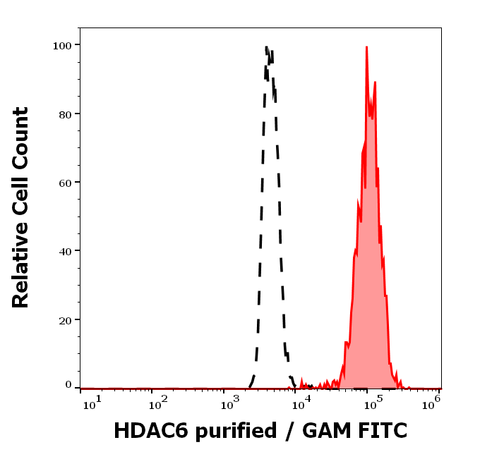
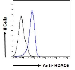
Separation of K562 cells stained using anti-human HDAC6 (236) purified antibody (concentration in sample 5.33 μg/ml, GAM APC, red-filled) from K562 cells unstained by primary antibody (GAM APC, black-dashed) in flow cytometry analysis (surface staining).
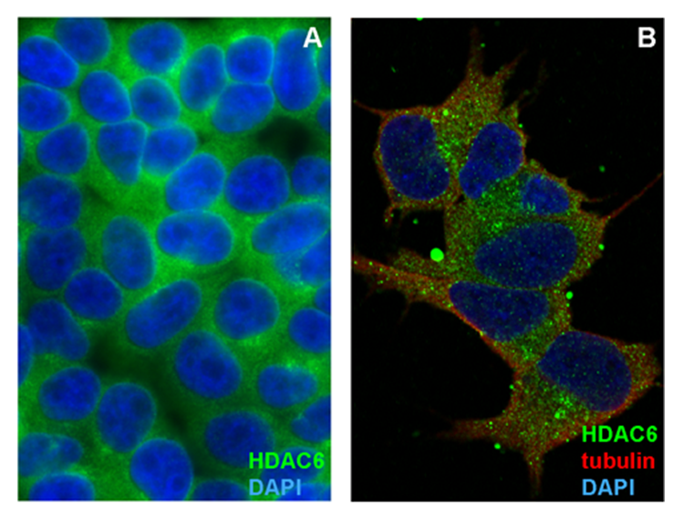
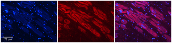
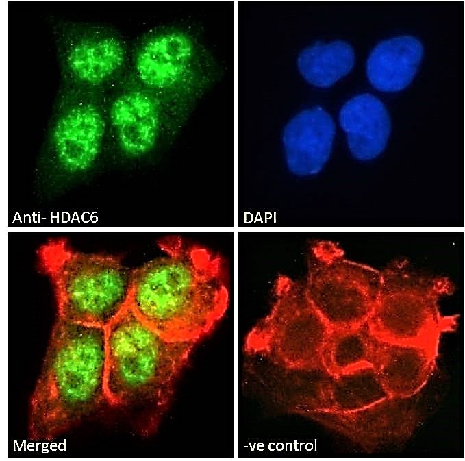
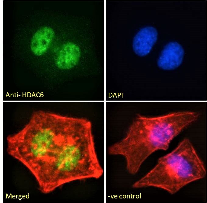
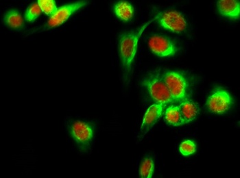
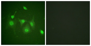
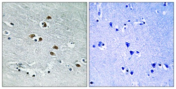
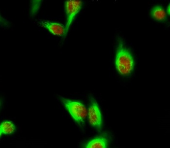
Immunocytochemistry staining of HDAC6 in formaldehyde-fixed and Triton-permeabilized HEK-293T cells (A) and SH-SY5Y cells (B) by mouse monoclonal antibody 236, followed by anti-mouse Alexa Fluor 488 (green), DNA indicated by DAPI (blue). In SH-SY5Y cells tubulin was stained by a rabbit polyclonal antibody, followed by anti-rabbit Alexa Fluor 647 (red, colocalization yellow).
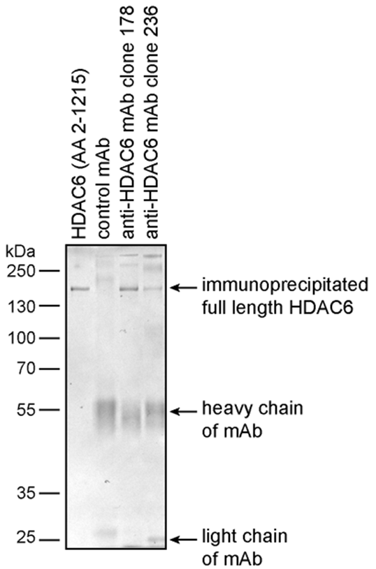
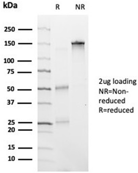
Immunoprecipitation of recombinant human HDAC6 by mouse monoclonal antibodies 178 and 236 using protein G-coated Dynabeads. Eluted proteins were processed by SDS PAAGE under reducing conditions and visualized by Coomassie Brilliant Blue.
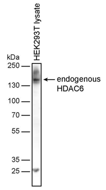
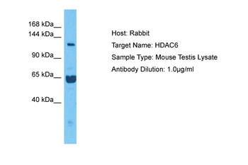
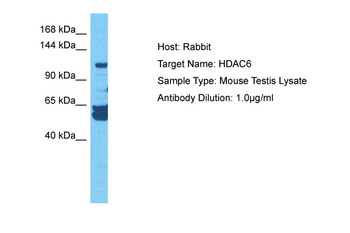
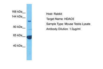
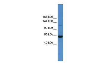
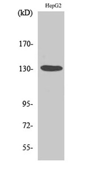
Western blotting analysis of human HDAC6 by mouse monoclonal antibody 236 in HEK-293T cell line under reducing conditions. PVDF membrane was blocked in 5% milk, and incubated with primary antibody (1 µg/ml), followed by HRP-conjugated anti-mouse secondary antibody.
Documents Download
Datasheet
Product Information
Request a Document
Protocol Information
WB
Western Blot (IB, immunoblot)
FC
Flow Cytometry
ICC
Immunocytochemistry
ELISA
Enzyme-linked Immunosorbent Assay (EIA)
IP
Immunoprecipitation
HDAC6 Antibody (orb699854)

- 0.0
Based on 0 reviews
Participating in our Biorbyt product reviews program enables you to support fellow scientists by sharing your firsthand experience with our products.
Login to Submit a ReviewAvailable Sizes
Select a size below
Free Secondary Antibody (20 ul)0/0
Please add an antibody product to your cart first.