You have no items in your shopping cart.
Description
Research Area
Cancer Biology; Musculoskeletal & Connective Tissue Research
Images & Validation
−Item 1 of 2
| Tested Applications | WB |
|---|---|
| Dilution Range | Western blot, 0.02-0.1 μg/ml |
| Reactivity | Gallus, Human, Monkey, Mouse, Rat, Zebrafish |
Key Properties
−| Antibody Type | Primary Antibody |
|---|---|
| Host | Rabbit |
| Clonality | Polyclonal |
| Isotype | IgG |
| Immunogen | HRP-Beta-Actin antibody was raised against a synthetic peptide containing 16 amino acids near the amino terminus of beta-actin. |
| Target | Actin, cytoplasmic 1 |
| Molecular Weight | 42 kDa |
| Purification | HRP-Beta-Actin antibody is affinity chromatography purified via peptide column. |
| Conjugation | HRP |
Storage & Handling
−| Storage | HRP-Beta-Actin antibody can be stored at 4°C for three months and -20°C, stable for up to one year. |
|---|---|
| Form/Appearance | Liquid |
| Buffer/Preservatives | Each vial contains 50% glycerol, 0.9% NaCl, 0.2% Na2HPO4. |
| Concentration | 0.5 mg/mL |
| Expiration Date | 12 months from date of receipt. |
| Disclaimer | For research use only |
Alternative Names
−ACTB; Beta-actin; B actin; Beta actin; PS1TP5BP1; β actin; BRWS1
Similar Products
−- Item 1 of 1
Beta-Actin Rabbit Polyclonal Antibody (HRP) [orb110039]
WB
Canine, Feline, Fish, Gallus, Guinea pig, Hamster, Insect, Porcine, Rabbit, Sheep
Human, Mouse, Rat
Rabbit
Polyclonal
HRP
100 μl 
Phospho-beta Actin (Tyr53) Rabbit Polyclonal Antibody (HRP) [orb503942]
IHC-Fr, IHC-P, WB
Bovine, Gallus, Porcine, Rabbit
Human, Mouse, Rat
Rabbit
Polyclonal
HRP
100 μlZebrafish Beta Actin/ACTB Rabbit Polyclonal Antibody (HRP) [orb2805628]
Zebrafish
Rabbit
Polyclonal
HRP
100 μgbeta Actin/ACTB Rabbit Polyclonal Antibody (HRP) [orb2576590]
Gallus, Human, Monkey, Mouse, Rat, Zebrafish
Rabbit
Polyclonal
HRP
100 μg

Quality Guarantee
Explore bioreagents carefree to elevate your research. All our products are rigorously tested for performance. If a product does not perform as described on its datasheet, our scientific support team will provide expert troubleshooting, a prompt replacement, or a refund. For full details, please see our Terms & Conditions and Buying Guide. Contact us at [email protected].
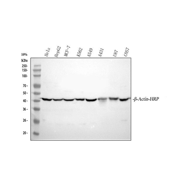
Western blot analysis of beta-Actin using anti-beta-Actin antibody. Electrophoresis was performed on a 5-20% SDS-PAGE gel at 70V (Stacking gel) / 90V (Resolving gel) for 2-3 hours. The sample well of each lane was loaded with 30 ug of sample under reducing conditions. Lane 1: human Hela whole cell lysates, Lane 2: human HepG2 whole cell lysates, Lane 3: human MCF-7 whole cell lysates, Lane 4: human K562 whole cell lysates, Lane 5: human A549 whole cell lysates, Lane 6: human A431 whole cell lysates, Lane 7: human U87 whole cell lysates, Lane 8: monkey COS-7 whole cell lysates. After electrophoresis, proteins were transferred to a nitrocellulose membrane at 150 mA for 50-90 minutes. Blocked the membrane with 5% non-fat milk/TBS for 1.5 hour at RT. The membrane was incubated with rabbit anti-beta-Actin antigen affinity purified polyclonal antibody at 0.1 µg/mL overnight at 4°C, then washed with TBS-0.1% Tween 3 times with 5 minutes each. The signal is developed using an Enhanced Chemiluminescent detection (ECL) kit with Tanon 5200 system. A specific band was detected for beta-Actin at approximately 42 kDa. The expected band size for beta-Actin is at 42 kDa.
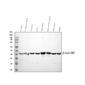
Western blot analysis of beta-Actin using anti-beta-Actin antibody. Electrophoresis was performed on a 5-20% SDS-PAGE gel at 70V (Stacking gel) / 90V (Resolving gel) for 2-3 hours. The sample well of each lane was loaded with 30 ug of sample under reducing conditions. Lane 1: rat heart tissue lysates, Lane 2: rat skeletal muscle tissue lysates, Lane 3: rat brain tissue lysates, Lane 4: rat PC-12 whole cell lysates, Lane 5: mouse heart tissue lysates, Lane 6: mouse skeletal tissue lysates, Lane 7: mouse brain tissue lysates, Lane 8: mouse NIH/3T3 whole cell lysates. After electrophoresis, proteins were transferred to a nitrocellulose membrane at 150 mA for 50-90 minutes. Blocked the membrane with 5% non-fat milk/TBS for 1.5 hour at RT. The membrane was incubated with rabbit anti-beta-Actin antigen affinity purified polyclonal antibody at 0.1 µg/mL overnight at 4°C, then washed with TBS-0.1% Tween 3 times with 5 minutes each. The signal is developed using an Enhanced Chemiluminescent detection (ECL) kit with Tanon 5200 system. A specific band was detected for beta-Actin at approximately 42 kDa. The expected band size for beta-Actin is at 42 kDa.
Quick Database Links
Gene Symbol
Actin, cytoplasmic 1
UniProt
UniProt Details
− No UniProt data available
Documents Download
Datasheet
Product Information
Request a Document
beta-Actin ACTB Rabbit Polyclonal Antibody (HRP) (orb1804654)

- 0.0
Based on 0 reviews
Participating in our Biorbyt product reviews program enables you to support fellow scientists by sharing your firsthand experience with our products.
Login to Submit a ReviewAvailable Sizes
Select a size below
Free Secondary Antibody (20 ul)0/0
Please add an antibody product to your cart first.