You have no items in your shopping cart.
Description
Research Area
Cell Biology
Images & Validation
−Item 1 of 2
| Tested Applications | WB |
|---|---|
| Reactivity | Human, Mouse, Plant, Porcine |
| Application Notes |
Key Properties
−| Antibody Type | Primary Antibody |
|---|---|
| Clonality | Monoclonal |
| Isotype | Mouse IgM |
| Clone No. | TU-02 |
| Immunogen | microtubule proteins from porcine brain |
| Target | alpha-Tubulin |
| Purification | Purified by sequential steps of physicochemical fractionation (differential precipitation and solid-phase chromatography methods). |
| Conjugation | Unconjugated |
Storage & Handling
−| Storage | Maintain refrigerated at 2-8°C for up to 2 weeks. For long term storage store at -20°C in small aliquots to prevent freeze-thaw cycles. |
|---|---|
| Buffer/Preservatives | Tris buffered saline (TBS), pH 8.0, 15 mM sodium azide |
| Concentration | 1 mg/ml |
| Expiration Date | 12 months from date of receipt. |
| Disclaimer | For research use only |
Alternative Names
−TUBA
Similar Products
−- Item 1 of 23
TUBA1B Antibody [orb344425]
ELISA, IF, IHC, Multiplex Assay, WB
Human
Mouse
Monoclonal
Unconjugated
100 μg - Item 1 of 23
TUBA1B Antibody [orb344426]
ELISA, IF, IHC, Multiplex Assay, WB
Human
Mouse
Monoclonal
Unconjugated
25 μl - Item 1 of 11
Tubulin alpha Mouse Monoclonal Antibody [orb738419]
FC, ICC, IF, IHC, WB
Human, Mouse, Rat
Mouse
Monoclonal
Unconjugated
100 μg - Item 1 of 9
alpha-Tubulin Antibody [orb44529]
FC, ICC, IHC-P, IP, WB
Aves, Human, Invertebrate, Mouse, Paramecium, Plant, Porcine, Yeast
Monoclonal
Unconjugated
0.1 mg - Item 1 of 6
alpha-Tubulin Antibody [orb44543]
ELISA, ICC, IHC-P, IP, WB
Canine, Human, Mouse, Plant, Porcine, Rat
Monoclonal
Unconjugated
0.1 mg


Quality Guarantee
Explore bioreagents carefree to elevate your research. All our products are rigorously tested for performance. If a product does not perform as described on its datasheet, our scientific support team will provide expert troubleshooting, a prompt replacement, or a refund. For full details, please see our Terms & Conditions and Buying Guide. Contact us at [email protected].
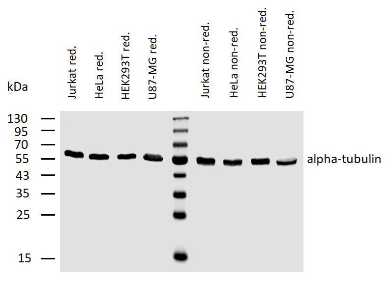
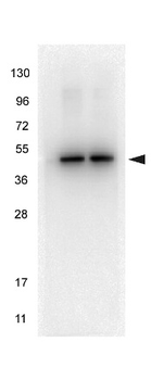
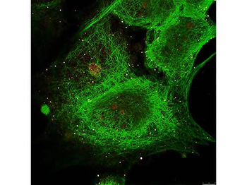
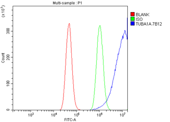
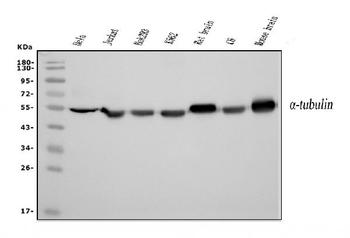
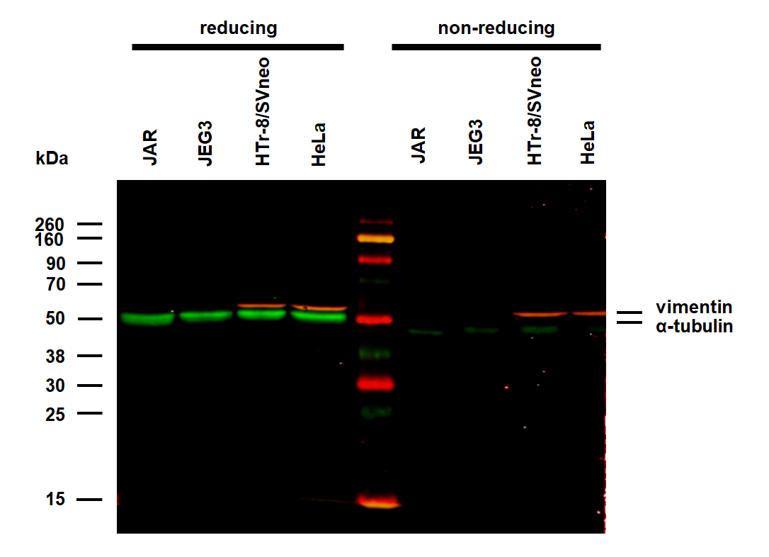
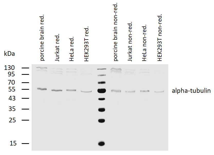
Western blotting analysis of human alpha-tubulin using mouse monoclonal antibody TU-02 on lysates (50 mM TRIS-Cl pH 6.8, 4M UREA, 4% SDS) of various cell lines under reducing and non-reducing conditions. Nitrocellulose membrane was probed with 2 µg/ml of mouse anti-alpha-tubulin monoclonal antibody followed by IRDye800-conjugated anti-mouse secondary antibody. A specific band was detected for alpha-tubulin at approximately 55 kDa.
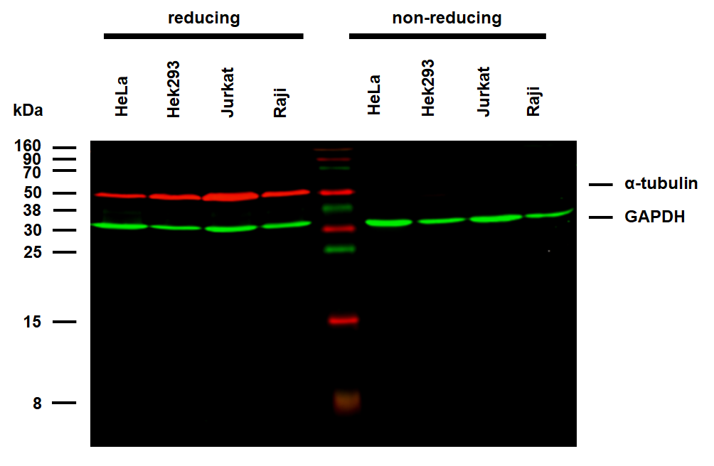
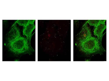
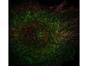
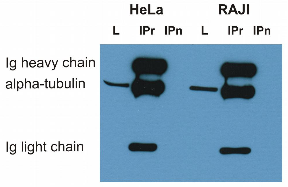
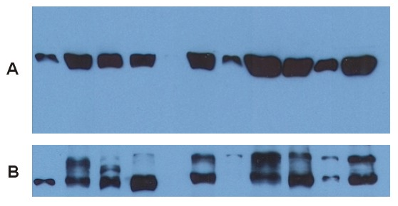
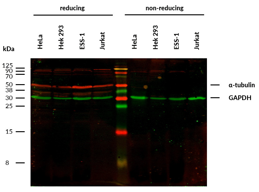
Anti-alpha-Tubulin Purified (TU-02) works in WB application under reducing conditions on RIPA cell extracts. Western blotting analysis was performed on whole cell extracts (RIPA lysis buffer) of HeLa, HEK 293, Jurkat, and Raji cell lines mixed and heated (100°C, 5 min) with reducing (2-mercaptoethanol) or non-reducing SDS-loading buffer. Samples were resolved using 15% Tris-glycine SDS gel electrophoresis. Nitrocellulose membrane blot was probed simultaneously with mouse IgM monoclonal antibody TU-02 (1 µg/ml) and anti-GAPDH mouse IgG1 monoclonal antibody FF26A (1 µg/ml) used as the loading control. Subclass-specific secondary antibodies IRDye 680RD Goat-anti-Mouse IgM (red) and IRDye 800CW Goat-anti-Mouse IgG (green) were used for multiplex fluorescent Western blot detection. Alpha-tubulin was detected at ~50 kDa in all tested cell lines. Anti-alpha-Tubulin Purified (TU-02) is not suitable for use in non-reducing conditions on RIPA cell extracts.
Documents Download
Datasheet
Product Information
Request a Document
alpha-Tubulin Antibody (orb44534)

- 0.0
Based on 0 reviews
Participating in our Biorbyt product reviews program enables you to support fellow scientists by sharing your firsthand experience with our products.
Login to Submit a ReviewAvailable Sizes
Select a size below
Free Secondary Antibody (20 ul)0/0
Please add an antibody product to your cart first.