You have no items in your shopping cart.
Featured
Description
Research Area
Neuroscience
Images & Validation
−Item 1 of 3
| Tested Applications | In vitro, In vivo, SDS-PAGE, WB |
|---|---|
| Application Notes |
Key Properties
−| Source | Recombinant |
|---|---|
| Expression System | E. coli |
| Biological Origin | Mouse |
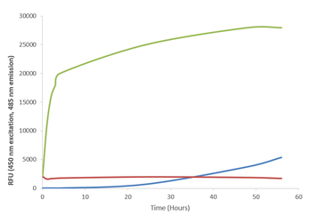
| Biological Activity | 100 µM alpha synuclein protein monomer seeded with 10 uM alpha synuclein protein PFF in 25 µM Thioflavin T (PBS pH 7.4, 100 µl reaction volume) generated an increased fluorescence intensity after incubation at 37°C with shaking at 600 rpm. Fluorescence was measured by excitation at 450 nm and emission at 485 nm on a Molecular Devices Gemini XPS microplate reader. |
| Target | Alpha Synuclein Monomers |
| Reactivity | Mouse |
| Tag | No tag |
| Protein Length | Full Length |
| Purification | Ion-exchange Purified |
| MW | ~14.46 kDa |
| Purity | >95% |
| Protein Sequence | MDVFMKGLSK AKEGVVAAAE KTKQGVAEAA GKTKEGVLYV GSKTKEGVVH GVTTVAEKTK EQVTNVGGAV VTGVTAVAQK TVEGAGNIAA ATGFVKKDQM GKGEEGYPQE GILEDMPVDP GSEAYEMPSE EGYQDYEPEA |
Storage & Handling
−| Storage | -80°C |
|---|---|
| Buffer/Preservatives | PBS pH 7.4 |
| Concentration | 2 mg/ml or 5 mg/ml |
| Expiration Date | 6 months from date of receipt. |
| Dry Ice Shipping | Please note: This product requires shipment on dry ice. A dry ice surcharge will apply. |
| Disclaimer | For research use only |
Alternative Names
−Alpha synuclein monomer, Alpha-synuclein monomer, Alpha synuclein protein monomer, Alpha synuclein monomer, Alpha-synuclein protein, Non-A beta component of AD amyloid protein, Non-A4 component of amyloid precursor protein, NACP protein, SNCA protein, NACP protein, PARK1 protein, Alpha synuclein monomers, SYN protein, Parkinson's disease familial 1 Protein
Similar Products
−- Item 1 of 5
Alpha Synuclein Monomers [orb1822364]
In vitro, In vivo, SDS-PAGE, WB
>95%
~14.46 kDa
Recombinant
100 μg - Item 1 of 2
Alpha Synuclein A53T Mutant Monomers [orb1822355]
In vitro, In vivo, SDS-PAGE, WB
>95%
~14.46 kDa
Recombinant
100 μg - Item 1 of 2
Alpha Synuclein Monomers [orb1822363]
In vitro, In vivo, SDS-PAGE, WB
>95%
~14.46 kDa
Recombinant
100 μg
Quality Guarantee
Explore bioreagents carefree to elevate your research. All our products are rigorously tested for performance. If a product does not perform as described on its datasheet, our scientific support team will provide expert troubleshooting, a prompt replacement, or a refund. For full details, please see our Terms & Conditions and Buying Guide. Contact us at [email protected].
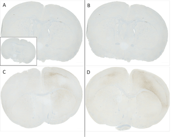
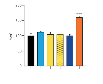
C57/BL6 mice were injected with sonicated recombinant mouse alpha synuclein monomers or fibrils at 8 weeks of age. Mice were unilaterally injected in the dorsal striatum (bregma AP + 0.2 mm, L +/1 2.0 mm, V - 3.0 mm) and sacrificed 30 days post-injection. (A) 1.25 uL mouse alpha synuclein monomers. (B) 2.5 uL mouse alpha synuclein monomers. (C) 2.5 ug alpha synuclein PFFs. (C) 5 ug alpha synuclein PFFs Inset: PBS (negative control). Primary antibody: Anti-Alpha Synuclein pSer129 at 1:10 000. Secondary antibody: anti-rabbit HRP. Mice injected with PFF displayed alpha synuclein staining in the striatum and cortex and contralateral to the injection site.
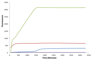
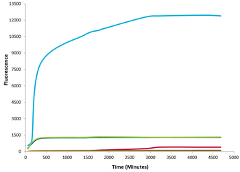
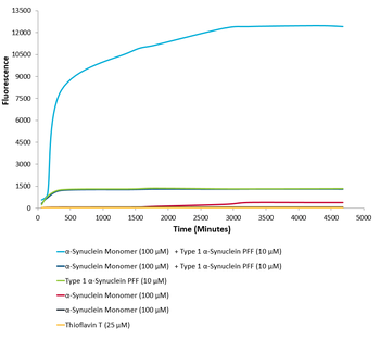
Type 1 alpha synuclein pre-formed fibrils seed the formation of new alpha synuclein fibrils from the pool of alpha synuclein monomers. Thioflavin T is a fluorescent dye that binds to beta sheet-rich structures, such as those in alpha synuclein fibrils. Upon binding, the emission spectrum of the dye experiences a red-shift, and increased fluorescence intensity. Thioflavin T emission curves show increased fluorescence (correlated to alpha synuclein protein aggregation) over time when 10 μM of Type 1 alpha synuclein pre-formed fibrils is combined with 100 μM of alpha synuclein monomer, as compared to Type 1 alpha synuclein pre-formed fibrils or alpha synuclein monomer alone. Thioflavin T ex = 450 nm, em = 485 nm.
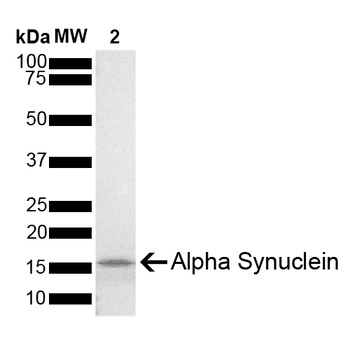
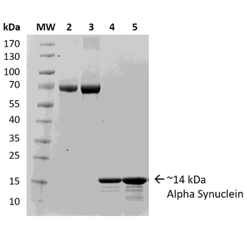
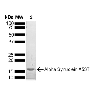
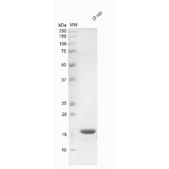
SDS-PAGE of ~14 kDa Mouse Recombinant Alpha Synuclein Protein Monomer. Lane 1: Molecular Weight Ladder (MW). Lane 2: Alpha Synuclein Protein Monomer (2 μg).
Quick Database Links
Gene Symbol
Alpha Synuclein Monomers
UniProt
UniProt Details
− No UniProt data available
Documents Download
Datasheet
Product Information
Request a Document
Protocol Information
Protein Handling and Storage Guide
Protein Handling Guide
WB
Western Blot (IB, immunoblot)
SDS-PAGE
Sodium Dodecyl Sulphate PolyAcrylamide Gel Electrophoresis
Alpha Synuclein Monomers (orb1822361)

- 0.0
Based on 0 reviews
Participating in our Biorbyt product reviews program enables you to support fellow scientists by sharing your firsthand experience with our products.
Login to Submit a ReviewAvailable Sizes
Select a size below