You have no items in your shopping cart.
Description
Research Area
Metabolism Research
Images & Validation
−Item 1 of 4
| Tested Applications | ELISA, IP, WB |
|---|---|
| Dilution Range | ELISA: 1:7,000, IP: 1:100, WB: 1:1,000 - 1:5,000 |
| Reactivity | Human, Rabbit |
| Application Notes |
Key Properties
−| Antibody Type | Primary Antibody |
|---|---|
| Host | Goat |
| Clonality | Polyclonal |
| Isotype | IgG |
| Immunogen | Aldolase [Rabbit Muscle] |
| Target | ALDOA |
| Purity | Anti-ALDOLASE was prepared from monospecific antiserum by a delipidation, salt fractionation and ion exchange chromatography. Assay by immunoelectrophoresis resulted in a single precipitin arc against anti-Goat Serum, purified and partially purified Aldolase [Rabbit Muscle]. |
| Conjugation | Unconjugated |
Storage & Handling
−| Storage | Store vial at -20° C or below prior to opening. This vial contains a relatively low volume of reagent (25 µL). To minimize loss of volume dilute 1:10 by adding 225 µL of the buffer stated above directly to the vial. Recap, mix thoroughly and briefly centrifuge to collect the volume at the bottom of the vial. Use this intermediate dilution when calculating final dilutions as recommended below. Store the vial at -20°C or below after dilution. Avoid cycles of freezing and thawing. |
|---|---|
| Form/Appearance | Liquid (sterile filtered) |
| Buffer/Preservatives | Preservative: 0.01% (w/v) Sodium Azide. Stabilizer: None; Buffer: 0.02 M Potassium Phosphate, 0.15 M Sodium Chloride, pH 7.2 |
| Concentration | 1 mg/ml |
| Expiration Date | 12 months from date of receipt. |
| Dry Ice Shipping | Please note: This product requires shipment on dry ice. A dry ice surcharge will apply. |
| Disclaimer | For research use only |
Alternative Names
−goat anti-Aldolase Antibody, Fructose-bisphosphate aldolase A, Muscle-type aldolase
Similar Products
−- Item 1 of 8
Aldolase/ALDOA Rabbit Polyclonal Antibody [orb570364]
ELISA, FC, ICC, IF, IHC, WB
Human, Mouse, Rat
Rabbit
Polyclonal
Unconjugated
100 μg - Item 1 of 6
Aldolase/ALDOA Mouse Monoclonal Antibody [orb692226]
FC, ICC, IF, IHC, WB
Human
Mouse
Monoclonal
Unconjugated
100 μg - Item 1 of 1
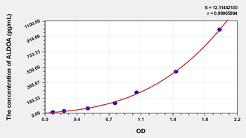
Human Aldolase A, Fructose Bisphosphate (ALDOA) ELISA Kit [orb778557]
Human
15.63-1000 pg/mL
6.9 pg/mL
96 T, 48 T - Item 1 of 1
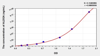
Rat Aldolase A, Fructose Bisphosphate (ALDOA) ELISA Kit [orb777216]
Rat
0.16-10 ng/mL
0.063 ng/mL
96 T, 48 T - Item 1 of 3
ALDOA Antibody (C-term) [orb1931515]
IHC-P, WB
Rabbit
Human, Mouse
Rabbit
Polyclonal
Unconjugated
50 μl, 100 μl

Quality Guarantee
Explore bioreagents carefree to elevate your research. All our products are rigorously tested for performance. If a product does not perform as described on its datasheet, our scientific support team will provide expert troubleshooting, a prompt replacement, or a refund. For full details, please see our Terms & Conditions and Buying Guide. Contact us at [email protected].
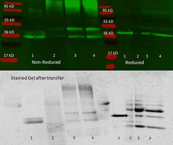
Anti aldolase antibody – Immunoprecipitation and Western Blot. 300 µl aliquots of whole anti-aldolase antiserum (orb750415) were used to precipitate varying amounts of purified aldolase and precipitates with controls were compared by SDS-PAGE and Western blot. Samples shown in the image are: 1. Purified aldolase 2. 300 µl antiserum with no antigen (negative control) 3. 300 µl antiserum with ~100 µl aldolase (2.5 mg/ml) 4. 300 µl antiserum with ~200 µl aldolase (2.5 mg/ml) For the precipitation, 300 ul of antiserum and an equal volume of aldolase antigen in PBS was incubated ~24 hrs at 4°C, centrifuged for 6 minutes at 13K RPM, washed once with PBS, centrifuged and dissolved in 60 ul 0.1 N NaOH. 90 ul of PBS was added, the sample was divided in 2 portions, and an equal volume of reducing (+4% BME) or non-reducing 2X sample buffer was added. The reduced samples were boiled for five minutes, and all samples were run at 140 V for ~45 minutes on a 4-20% tris/glycine gradient gel. Gel was stained, destained and imaged (see attached) using standard protocols. Precipitation of aldolase was confirmed by comparison of increasing amounts of antigen with the control protein by SDS PAGE and observation of a 40-45 kD MW band corresponding to Aldolase. Additional higher and lower molecular weight bands correspond to serum proteins.
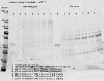
Anti aldolase antibody– Immunoprecipitation- Immunoprecipitation was performed with 300 ul of anti aldolase antiserum and an equal volume of varied amounts (diluted from a stock solution of ~2.5 mg/ml) of purified aldolase in PBS. Antibody/Antigen mixture was incubated ~24 hrs at 4°C, centrifuged for 6 minutes at 13K RPM, washed once with PBS, centrifuged and dissolved in 60 ul 0.1 N NaOH. 90 ul of PBS was added, the sample was divided in 2 portions, and an equal volume of reducing (+4% BME) or non-reducing 2X sample buffer was added. The reduced samples were boiled for five minutes, and all samples were run at 140 V for ~45 minutes on a 4-20% tris/glycine gradient gel. Gel was stained, destained and imaged (see attached) using standard protocols. Precipitation of aldolase was confirmed by comparison of increasing amounts of antigen with the control protein by SDS PAGE and observation of a 40-45 kD MW band corresponding to Aldolase. Additional higher and lower molecular weight bands correspond to serum proteins.
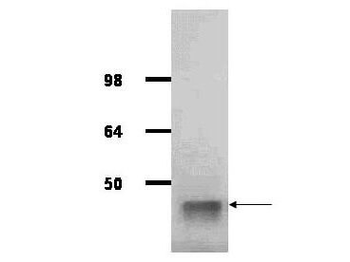
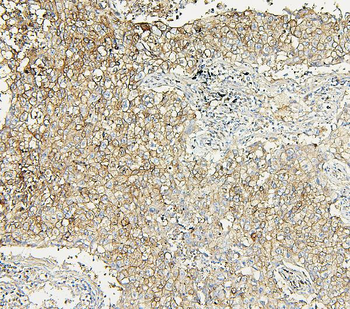
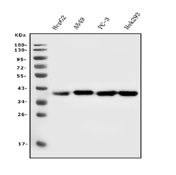
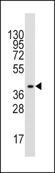
IgG purified antibody to rabbit muscle aldolase was used at a 1:1000 dilution to detect human aldolase by Western blot. A whole cell lysate prepared from human derived A293 cells was loaded on a 4-12% tris glycine gradient gel for SDS-PAGE. The gel was transferred to nitro-cellulose using standard techniques. Antibody reaction with the membrane occurred overnight at 4°C in TTBS supplemented with 2% non-fat dry milk. Color was allowed to develop using SuperSignal West Pico Chemiluminescent Substrate (PIERCE). Other detection methods will yield similar results. This antibody clearly detects a band at ~41 kDa consistent with human aldolase.

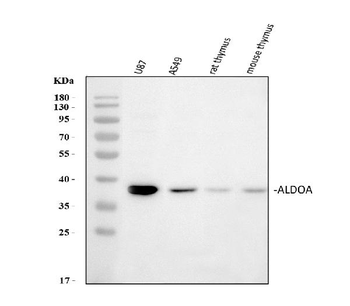
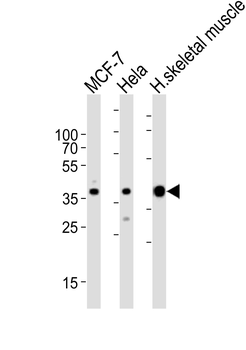
Western Blot of Goat anti-Aldolase Antibody. Lane 1: Hela lysate (p/n orb348668). Lane 2: HEK293 lysate (p/n orb348669). Lane 3: Jurkat lysate (p/n orb348674). Load: 25 µg per lane. Primary antibody: Goat anti-Aldolase Antibody at 1:1000 overnight at 4°C. Secondary antibody: HRP Dk-a-Gt IgG secondary antibody (p/n orb347031) at 1:40000 for 30 min at RT. Block: (p/n orb348637) for 30 min at RT. Predicted/Observed size: 39 kDa, 41 kDa for Aldolase.
Quick Database Links
UniProt Details
− No UniProt data available
NCBI Reference Sequences
−Associated Accession Numbers
Curated reference sequences for the gene transcript and protein product| Protein | NP_001075707.1 |
|---|
Documents Download
Datasheet
Product Information
Request a Document
Protocol Information
WB
Western Blot (IB, immunoblot)
ELISA
Enzyme-linked Immunosorbent Assay (EIA)
IP
Immunoprecipitation
ALDOA Antibody (orb344331)

- 0.0
Based on 0 reviews
Participating in our Biorbyt product reviews program enables you to support fellow scientists by sharing your firsthand experience with our products.
Login to Submit a ReviewAvailable Sizes
Select a size below
Free Secondary Antibody (20 ul)0/0
Please add an antibody product to your cart first.