You have no items in your shopping cart.
Featured
KO/KD
Validated
Validated
Description
Research Area
Neuroscience; Metabolism Research
Images & Validation
−Item 1 of 9
| Tested Applications | ICC, IHC, IP, KO/KD Validated, WB |
|---|---|
| Dilution Range | WB (1:1000); ICC/IF (1:200); IP (1:200); IHC (1:100) |
| Reactivity | Human, Mouse, Rat |
| Application Notes |
Key Properties
−| Host | Mouse |
|---|---|
| Clonality | Monoclonal |
| Isotype | IgG1 |
| Clone No. | 8A3 |
| Immunogen | Full length recombinant human VSP35 |
| Target | VPS35 |
| Molecular Weight | 92 kDa |
| Purification | Protein G Purified |
| Conjugation | RPE |
Storage & Handling
−| Storage | Conjugated antibodies should be stored according to the product label |
|---|---|
| Buffer/Preservatives | 95.46mM Phosphate, 2.48mM MES and 2mM EDTA |
| Concentration | 1 mg/ml |
| Expiration Date | 12 months from date of receipt. |
| Disclaimer | For research use only |
Alternative Names
−VPS35, VPS35 Retromer Complex Component, Vacuolar Protein Sorting-Associated Protein 35, Vacuolar Protein Sorting 35 Homolog, Vesicle Protein Sorting 35, MEM3, PARK17, FLJ10752, HVPS35, Maternal-Embryonic 3, TCCCTA00141
Similar Products
−- Item 1 of 8
VPS35 Antibody (RPE) [orb612768]
ICC, IF, IP, KO/KD Validated, WB
Human, Mouse, Rat
Mouse
Monoclonal
RPE
100 μg - Item 1 of 8
VPS35 Antibody (RPE) [orb612787]
ICC, IP, KO/KD Validated, WB
Human, Mouse, Rat
Mouse
Monoclonal
RPE
100 μg - Item 1 of 8
VPS35 Antibody (RPE) [orb612825]
ICC, IHC, IP, KO/KD Validated, WB
Human, Mouse, Rat
Mouse
Monoclonal
RPE
100 μg - Item 1 of 7
VPS35 Antibody (RPE) [orb612844]
IHC, IP, KO/KD Validated, WB
Human, Mouse, Rat
Mouse
Monoclonal
RPE
100 μg
Quality Guarantee
Explore bioreagents carefree to elevate your research. All our products are rigorously tested for performance. If a product does not perform as described on its datasheet, our scientific support team will provide expert troubleshooting, a prompt replacement, or a refund. For full details, please see our Terms & Conditions and Buying Guide. Contact us at [email protected].
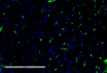
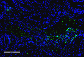
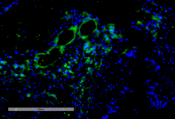
Immunohistochemistry analysis using Mouse Anti-VPS35 Monoclonal Antibody, Clone 8A3. Tissue: Kidney. Species: Mouse. Primary Antibody: Mouse Anti-VPS35 Monoclonal Antibody at 1:100 for Overnight at 4°C, then 30 min at 37°C. Secondary Antibody: Goat Anti-Mouse IgG (H+L): FITC for 45 min at 37°C. Counterstain: DAPI for 3 min at RT. Magnification: 20X.
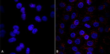
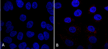
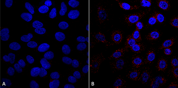
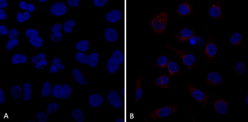
Immunocytochemistry/Immunofluorescence analysis using Mouse Anti-VPS35 Monoclonal Antibody, Clone 8A3. Tissue: A549 cells. Species: Human. Primary Antibody: Mouse Anti-VPS35 Monoclonal Antibody at 1:5 (tissue culture supernatant). Secondary Antibody: Donkey anti-mouse: Alexa Fluor 594 at 1:4000 in 0.2% BSA PBS. Counterstain: DAPI. Localization: Vesicles. A) VPS35 KO A549 cells B) WT A549 cells.
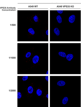
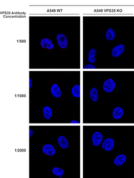
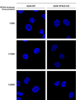
Immunocytochemistry/Immunofluorescence analysis using Mouse Anti-VPS35 Monoclonal Antibody, Clone 8A3. Tissue: A549 WT, VPS35 KO cells. Species: Human. Primary Antibody: Mouse Anti-VPS35 Monoclonal Antibody. Secondary Antibody: Donkey Anti-Mouse AlexaFluor 594. Clone can detect VPS35 at 1/2000 concentration.
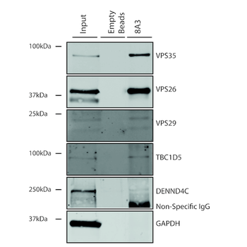
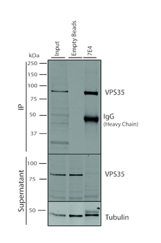
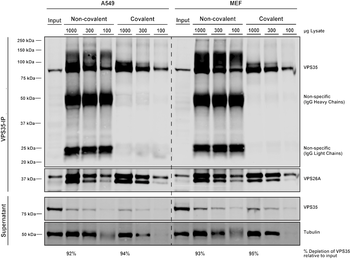
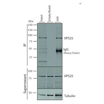
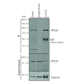
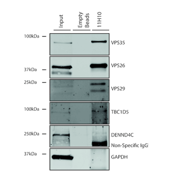
Immunoprecipitation analysis using Mouse Anti-VPS35 Monoclonal Antibody, Clone 8A3. Tissue: A549 cells. Species: Human. Primary Antibody: Mouse Anti-VPS35 Monoclonal Antibody.
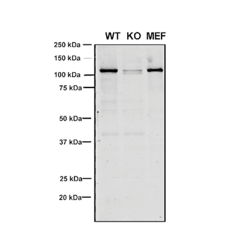
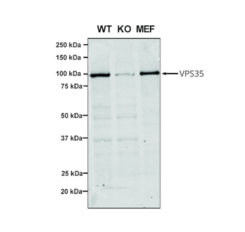
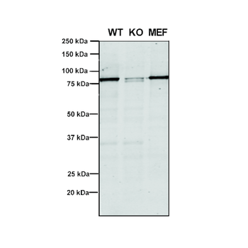
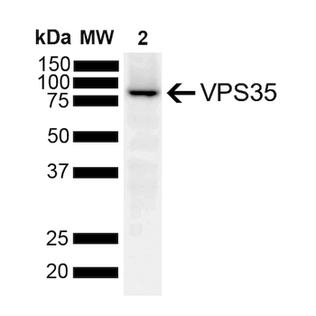
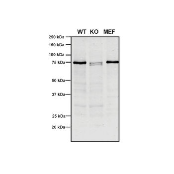
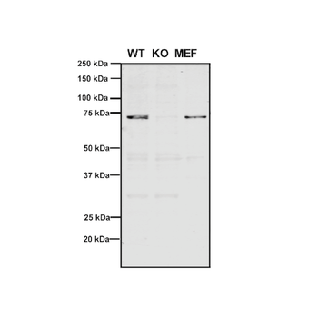
Western Blot analysis of Human, Mouse A549, MEF showing detection of VPS35 protein using Mouse Anti-VPS35 Monoclonal Antibody, Clone 8A3. Lane 1: Molecular Weight Ladder. Lane 2: VPS35 KO A549 cells. Lane 3: mouse embryonic fibroblast cells. Load: 8 μg each A549 and MEF. Primary Antibody: Mouse Anti-VPS35 Monoclonal Antibody at 1:5 (tissue culture supernatant). Secondary Antibody: Donkey anti-mouse IRDye 800CW at 1:25000 in TBS-T.
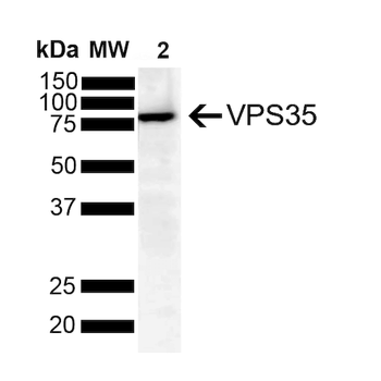
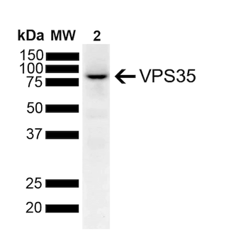
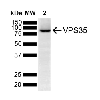
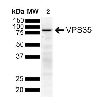
Western Blot analysis of Human SH-SY5Y showing detection of VPS35 protein using Mouse Anti-VPS35 Monoclonal Antibody, Clone 8A3. Lane 1: Molecular Weight Ladder. Lane 2: SH-SY5Y (10 ug). Load: 10 μg. Block: 5% Skim Milk powder in TBST. Primary Antibody: Mouse Anti-VPS35 Monoclonal Antibody at 1:1000 for 2 hours at RT with shaking. Secondary Antibody: Goat anti-mouse IgG:HRP at 1:4000 for 1 hour at RT with shaking. Color Development: Chemiluminescent for HRP (Moss) for 5 min in RT.
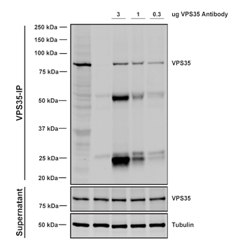
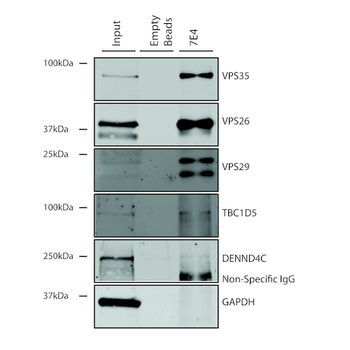
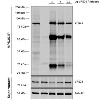
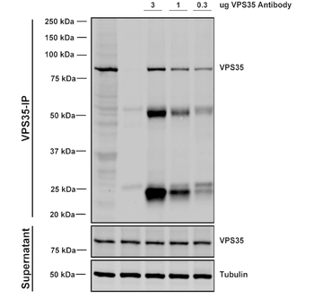
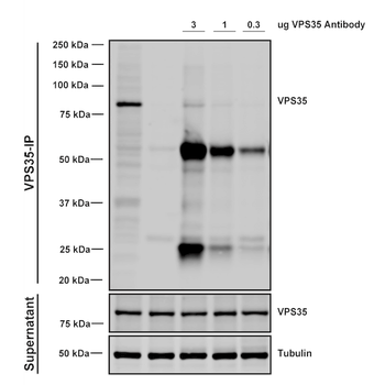
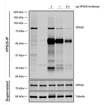
Immunoprecipitation analysis using Mouse Anti-VPS35 Monoclonal Antibody, Clone 8A3. Tissue: A549 cells. Species: Human. Primary Antibody: Mouse Anti-VPS35 Monoclonal Antibody. Three amounts of (3, 1 and 0.3 ug) were non-covalently coupled to 10uL of A/G sepharose beads for 1 hour at 4°C and next incubated with 250ug of A549 lysate for 2 hours at 4°C.
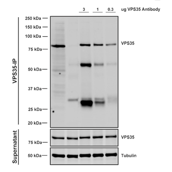
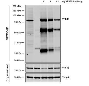
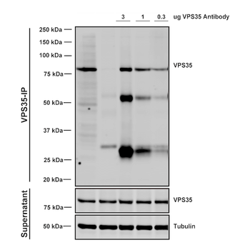
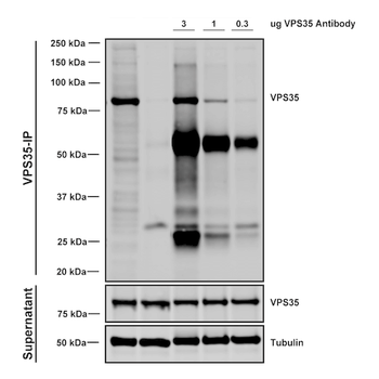
Immunoprecipitation analysis using Mouse Anti-VPS35 Monoclonal Antibody, Clone 8A3. Tissue: embryonic fibroblast. Species: Mouse. Primary Antibody: Mouse Anti-VPS35 Monoclonal Antibody. Three amounts of (3, 1 and 0.3 ug) were non-covalently coupled to 10uL of A/G sepharose beads for 1 hour at 4°C and next incubated with 250ug of MEF lysate for 2 hours at 4°C.
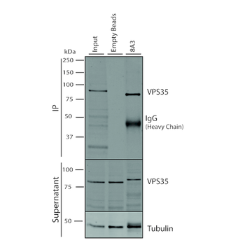
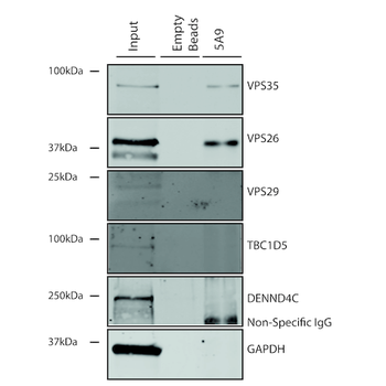
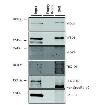
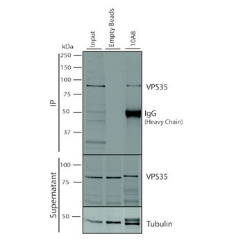
Immunoprecipitation analysis using Mouse Anti-VPS35 Monoclonal Antibody, Clone 8A3. Tissue: A549 cells. Species: Human. Primary Antibody: Mouse Anti-VPS35 Monoclonal Antibody. 500 μL cell culture supernatants were incubated with 10 μL of Protein A/G resin beads for 1 hour at 4°C.
Quick Database Links
UniProt Details
− No UniProt data available
NCBI Gene Details
− No NCBI Gene data available
NCBI Reference Sequences
−Associated Accession Numbers
Curated reference sequences for the gene transcript and protein product| Protein | NP_060676.2 |
|---|
Documents Download
Datasheet
Product Information
Request a Document
Protocol Information
WB
Western Blot (IB, immunoblot)
IHC
Immunohistochemistry
ICC
Immunocytochemistry
IP
Immunoprecipitation
VPS35 Antibody (RPE) (orb612806)

- 0.0
Based on 0 reviews
Participating in our Biorbyt product reviews program enables you to support fellow scientists by sharing your firsthand experience with our products.
Login to Submit a ReviewAvailable Sizes
Select a size below
Choose Conjugation or Carrier Free Version
Free Secondary Antibody (20 ul)0/0
Please add an antibody product to your cart first.