You have no items in your shopping cart.
Featured
KO/KD
Validated
Validated
Description
Research Area
Neuroscience
Images & Validation
−Item 1 of 8
| Tested Applications | ICC, IF, IP, KO/KD Validated, WB |
|---|---|
| Dilution Range | WB (1:1000); ICC/IF (1:200); IP (1:200) |
| Reactivity | Human, Mouse, Rat |
| Application Notes |
Key Properties
−| Host | Mouse |
|---|---|
| Clonality | Monoclonal |
| Isotype | IgG2a |
| Clone No. | 70000 |
| Immunogen | Full length recombinant human VSP35 |
| Target | VPS35 |
| Molecular Weight | 92 kDa |
| Purification | Protein G Purified |
| Conjugation | FITC |
Storage & Handling
−| Storage | Conjugated antibodies should be stored according to the product label |
|---|---|
| Buffer/Preservatives | 640.91mM DMSO, 136.36 mM Ethanolamine, 126.89 mM chlorides, 9.09mM phosphates, 9.09mM NaHCO3 |
| Concentration | 1 mg/ml |
| Expiration Date | 12 months from date of receipt. |
| Disclaimer | For research use only |
Alternative Names
−VPS35, VPS35 Retromer Complex Component, Vacuolar Protein Sorting-Associated Protein 35, Vacuolar Protein Sorting 35 Homolog, Vesicle Protein Sorting 35, MEM3, PARK17, FLJ10752, HVPS35, Maternal-Embryonic 3, TCCCTA00141
Similar Products
−- Item 1 of 9
VPS35 Antibody (FITC) [orb612802]
ICC, IHC, IP, KO/KD Validated, WB
Human, Mouse, Rat
Mouse
Monoclonal
FITC
100 μg - Item 1 of 8
VPS35 Antibody (FITC) [orb612783]
ICC, IP, KO/KD Validated, WB
Human, Mouse, Rat
Mouse
Monoclonal
FITC
100 μg - Item 1 of 8
VPS35 Antibody (FITC) [orb612821]
ICC, IHC, IP, KO/KD Validated, WB
Human, Mouse, Rat
Mouse
Monoclonal
FITC
100 μg - Item 1 of 7
VPS35 Antibody (FITC) [orb612840]
IHC, IP, KO/KD Validated, WB
Human, Mouse, Rat
Mouse
Monoclonal
FITC
100 μg 
VPS35 Rabbit Polyclonal Antibody (FITC) [orb464526]
IF
Bovine, Canine, Equine, Gallus, Porcine, Rabbit, Sheep
Human, Mouse, Rat
Rabbit
Polyclonal
FITC
100 μl

Quality Guarantee
Explore bioreagents carefree to elevate your research. All our products are rigorously tested for performance. If a product does not perform as described on its datasheet, our scientific support team will provide expert troubleshooting, a prompt replacement, or a refund. For full details, please see our Terms & Conditions and Buying Guide. Contact us at [email protected].
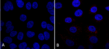
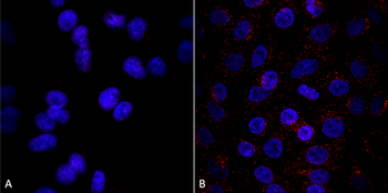
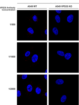
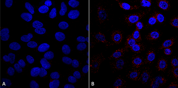
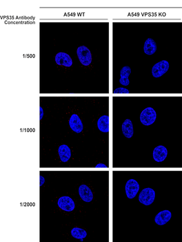
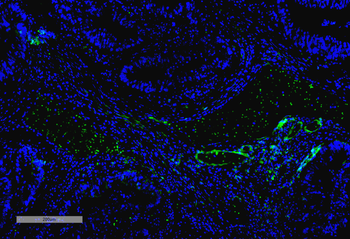
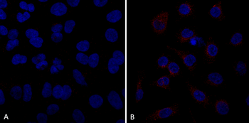
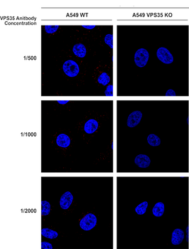
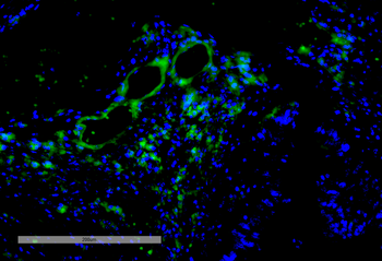
Immunocytochemistry/Immunofluorescence analysis using Mouse Anti-VPS35 Monoclonal Antibody, Clone 7E4. Tissue: A549 cells. Species: Human. Primary Antibody: Mouse Anti-VPS35 Monoclonal Antibody at 1:5 (tissue culture supernatant). Secondary Antibody: Donkey anti-mouse: Alexa Fluor 594 at 1:4000 in 0.2% BSA PBS. Counterstain: DAPI. Localization: Vesicles. A) VPS35 KO A549 cells B) WT A549 cells.
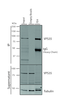
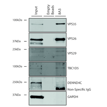
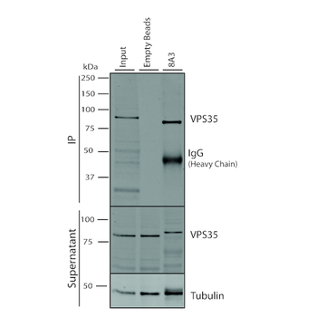
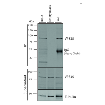
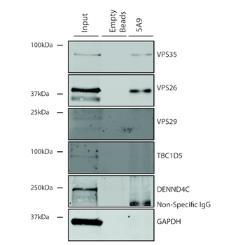
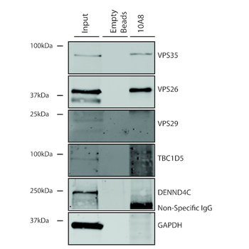
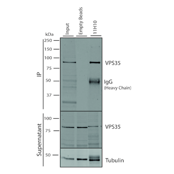
Immunoprecipitation analysis using Mouse Anti-VPS35 Monoclonal Antibody, Clone 7E4. Tissue: A549 cells. Species: Human. Primary Antibody: Mouse Anti-VPS35 Monoclonal Antibody. 500 μL cell culture supernatants were incubated with 10 μL of Protein A/G resin beads for 1 hour at 4°C. clone 7E4 depletes virtually all of the VPS35 from the A549 cell extract.
Immunoprecipitation analysis using Mouse Anti-VPS35 Monoclonal Antibody, Clone 7E4. Tissue: A549 cells. Species: Human. Primary Antibody: Mouse Anti-VPS35 Monoclonal Antibody.
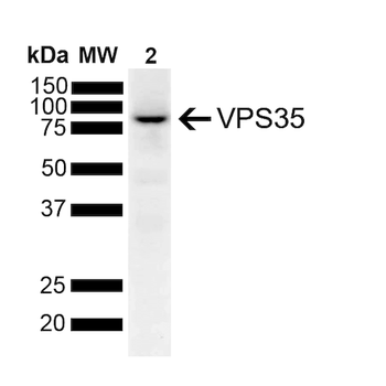
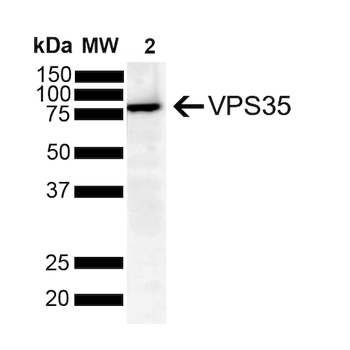
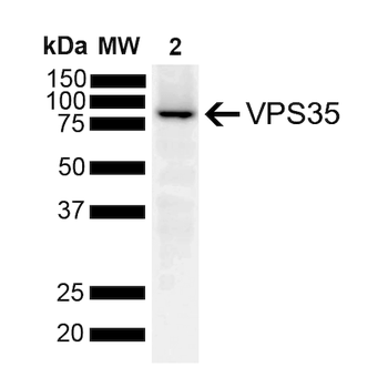
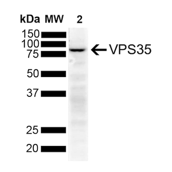
Western Blot analysis of Human SH-SY5Y lysates showing detection of 91.7 kDa VPS35 protein using Mouse Anti-VPS35 Monoclonal Antibody, Clone 7E4. Lane 1: Molecular Weight Ladder. Lane 2: SH-SY5Y. Load: 10 μg. Block: 5% Skim Milk powder in TBST. Primary Antibody: Mouse Anti-VPS35 Monoclonal Antibody at 1:1000 for 2 hours at RT with shaking. Secondary Antibody: Goat anti-mouse IgG:HRP at 1:4000 for 1 hour at RT with shaking. Color Development: Chemiluminescent for HRP (Moss) for 5 min in RT. Predicted/Observed Size: 91.7 kDa.
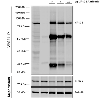
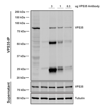
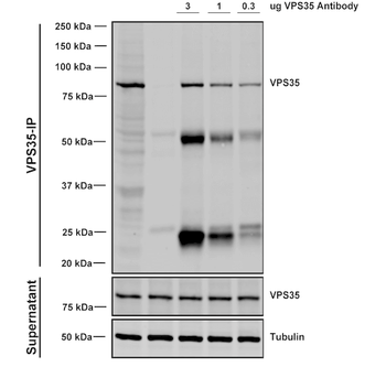
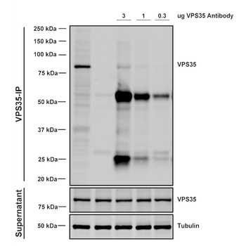
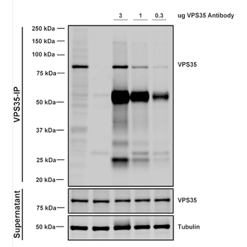
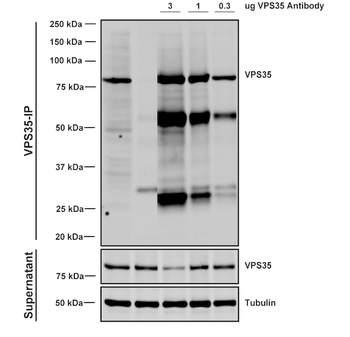
Immunoprecipitation analysis using Mouse Anti-VPS35 Monoclonal Antibody, Clone 7E4. Tissue: A549 cells. Species: Human. Primary Antibody: Mouse Anti-VPS35 Monoclonal Antibody. Three amounts of (3, 1 and 0.3 ug) were non-covalently coupled to 10uL of A/G sepharose beads for 1 hour at 4°C and next incubated with 250ug of A549 lysate for 2 hours at 4°C.
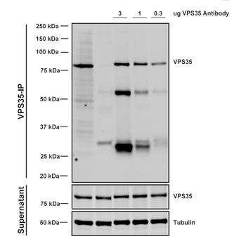
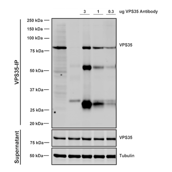
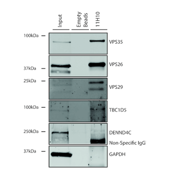
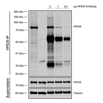
Immunoprecipitation analysis using Mouse Anti-VPS35 Monoclonal Antibody, Clone 7E4. Tissue: embryonic fibroblast. Species: Mouse. Primary Antibody: Mouse Anti-VPS35 Monoclonal Antibody. Three amounts of (3, 1 and 0.3 ug) were non-covalently coupled to 10uL of A/G sepharose beads for 1 hour at 4°C and next incubated with 250ug of MEF lysate for 2 hours at 4°C.
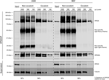
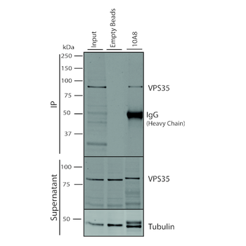
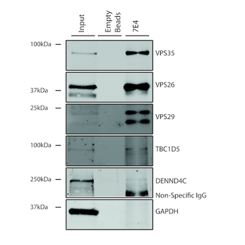
Immunoprecipitation analysis using Mouse Anti-VPS35 Monoclonal Antibody, Clone 7E4. Tissue: MEF, A549 cells. Species: Human, Mouse. Primary Antibody: Mouse Anti-VPS35 Monoclonal Antibody at 1:5 (tissue culture supernatant). 10 ug antibody were coupled to 10 uL A/G resin beads either covalently (with DMP) or non-covalently (1 hour at 4 degrees). The antibody immunoprecipitates VPS35 in mouse and human cells effectively when covalently coupled to the beads.
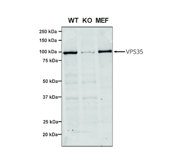
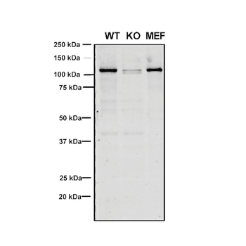
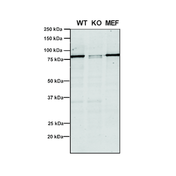
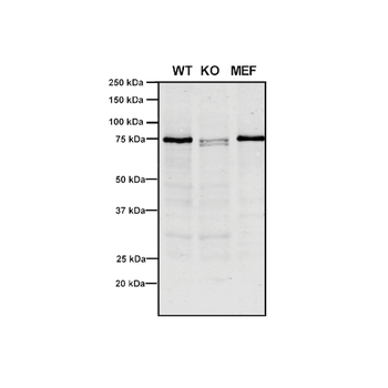
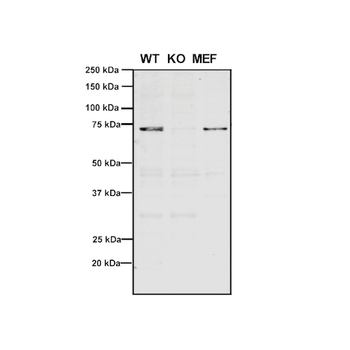
Western Blot analysis of Human, Mouse A549, MEF showing detection of VPS35 protein using Mouse Anti-VPS35 Monoclonal Antibody, Clone 7E4. Lane 1: Molecular Weight Ladder. Lane 2: VPS35 KO A549 cells. Lane 3: mouse embryonic fibroblast cells. Load: 8 μg each A549 and MEF. Primary Antibody: Mouse Anti-VPS35 Monoclonal Antibody at 1:5 (tissue culture supernatant). Secondary Antibody: Donkey anti-mouse IRDye 800CW at 1:25000 in TBS-T.
Quick Database Links
UniProt Details
− No UniProt data available
NCBI Gene Details
− No NCBI Gene data available
NCBI Reference Sequences
−Associated Accession Numbers
Curated reference sequences for the gene transcript and protein product| Protein | NP_060676.2 |
|---|
Documents Download
Datasheet
Product Information
Request a Document
Protocol Information
WB
Western Blot (IB, immunoblot)
IF
Immunofluorescence
ICC
Immunocytochemistry
IP
Immunoprecipitation
VPS35 Antibody (FITC) (orb612764)

- 0.0
Based on 0 reviews
Participating in our Biorbyt product reviews program enables you to support fellow scientists by sharing your firsthand experience with our products.
Login to Submit a ReviewAvailable Sizes
Select a size below
Choose Conjugation or Carrier Free Version
Free Secondary Antibody (20 ul)0/0
Please add an antibody product to your cart first.