You have no items in your shopping cart.
Description
Research Area
Cell Biology, Infectious Disease & Virology, Metabolism Research, Neuroscience, Signal Transduction
Images & Validation
−Item 1 of 4
| Tested Applications | FC, IF, IHC-P, WB |
|---|---|
| Dilution Range | IF: 1:25, WB: 1:4000, IHC-P: 1:25, FC: 1:25 |
| Reactivity | Human, Mouse, Rat |
| Predicted Reactivity | Canine |
Key Properties
−| Antibody Type | Primary Antibody |
|---|---|
| Host | Mouse |
| Clonality | Monoclonal |
| Isotype | IgG1,κ |
| Immunogen | This VCP antibody is generated from a mouse immunized with a recombinant protein of human VCP. Antigen Region: 400-806 aa. |
| Target | VCP |
| Molecular Weight | 89322 Da |
| Conjugation | Unconjugated |
Storage & Handling
−| Storage | Maintain refrigerated at 2-8°C for up to 2 weeks. For long term storage store at -20°C in small aliquots to prevent freeze-thaw cycles |
|---|---|
| Expiration Date | 12 months from date of receipt. |
| Disclaimer | For research use only |
Alternative Names
−Transitional endoplasmic reticulum ATPase, TER ATPase, 15S Mg(2+)-ATPase p97 subunit, Valosin-containing protein, VCP, VCP
Similar Products
−- Item 1 of 11
NVL Rabbit Polyclonal Antibody [orb1743797]
ELISA, FC, ICC, IF, IHC, WB
Human, Rat
Rabbit
Polyclonal
Unconjugated
100 μg - Item 1 of 10
VCP Rabbit Polyclonal Antibody [orb259631]
FC, ICC, IF, IHC, IHC-Fr, WB
Human, Mouse, Rat
Rabbit
Polyclonal
Unconjugated
100 μg - Item 1 of 8
VCP Rabbit Polyclonal Antibody [orb1289976]
ELISA, FC, IHC, WB
Human, Monkey, Mouse, Rat
Rabbit
Polyclonal
Unconjugated
100 μg - Item 1 of 8
VCP Rabbit Polyclonal Antibody [orb556833]
IHC, IHC-P, WB
Canine, Human, Mouse, Rat, Zebrafish
Rabbit
Polyclonal
Unconjugated
100 μl - Item 1 of 6
VCP Rabbit Polyclonal Antibody [orb107641]
FC, ICC, IF, IHC, IHC-Fr, WB
Human, Monkey, Mouse, Rat
Rabbit
Polyclonal
Unconjugated
100 μg
Quality Guarantee
Explore bioreagents carefree to elevate your research. All our products are rigorously tested for performance. If a product does not perform as described on its datasheet, our scientific support team will provide expert troubleshooting, a prompt replacement, or a refund. For full details, please see our Terms & Conditions and Buying Guide. Contact us at [email protected].
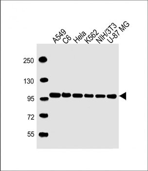
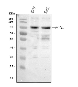
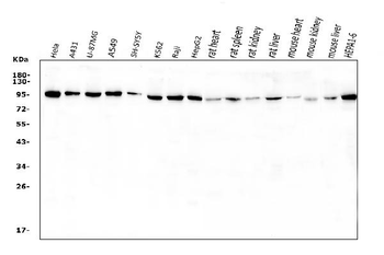
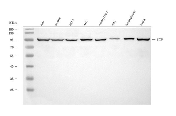
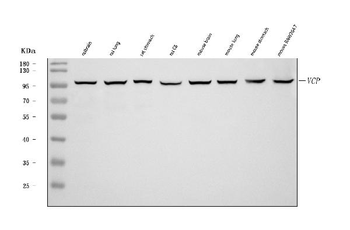
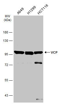
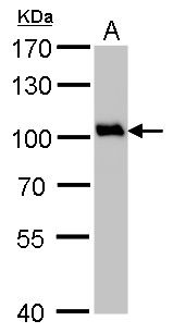
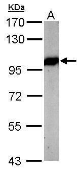
All lanes: Anti-VCP Antibody at1:4000 dilution. Lane 1: A549 whole cell lysate. Lane 2: C6 whole cell lysate. Lane 3: Hela whole cell lysate. Lane 4: K562 whole cell lysate. Lane 5: NIH/3T3 whole cell lysate. Lane 6: U-87 MG whole cell lysate. Lysates/proteins at 20 µg per lane. Secondary Goat Anti-mouse IgG, (H+L), Peroxidase conjugated at 1/10000 dilution. Predicted band size: 89 kDa. Blocking/Dilution buffer: 5% NFDM/TBST.
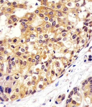
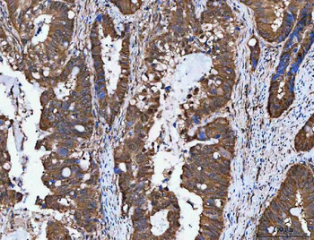
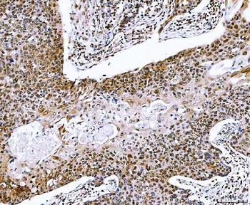
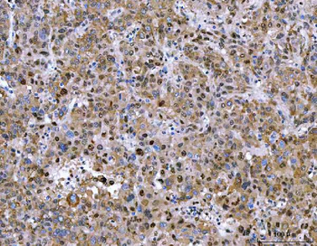
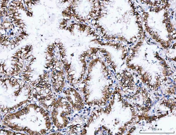
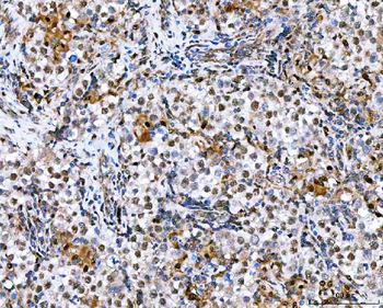
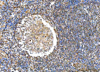
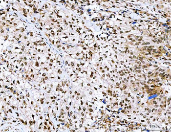
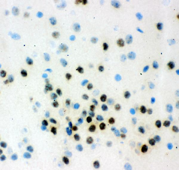
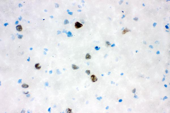
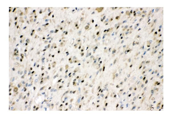
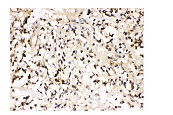
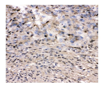
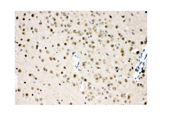
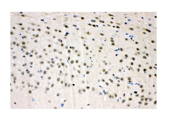
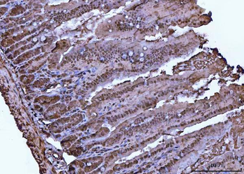
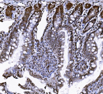
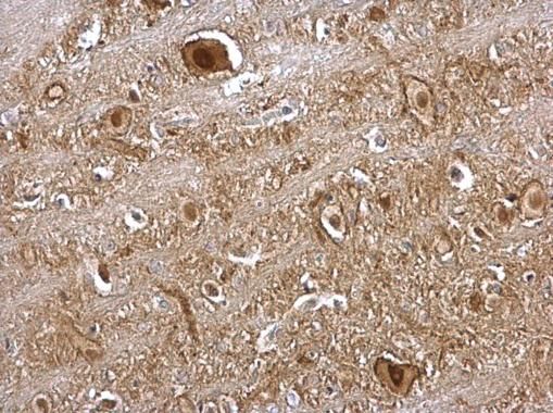
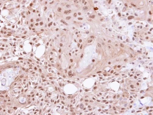
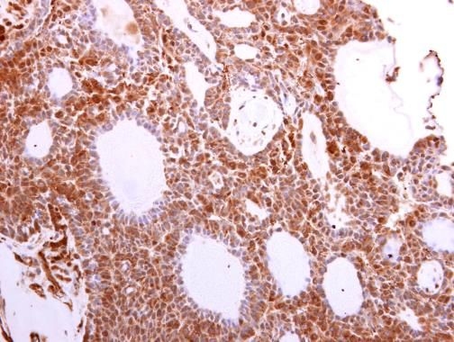
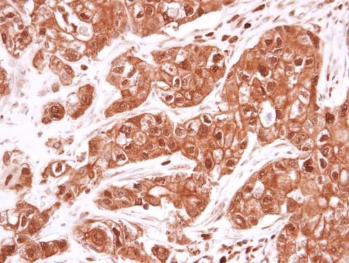
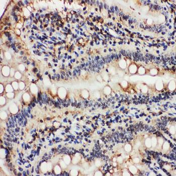
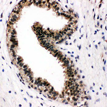
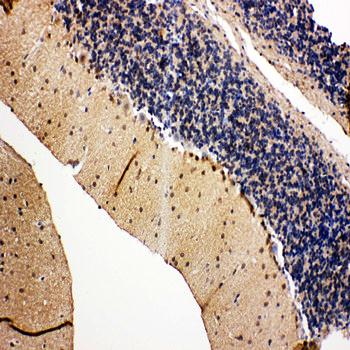
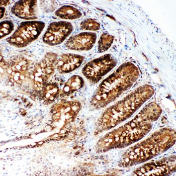
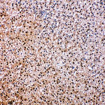
Staining VCP in human breast carcinoma sections by Immunohistochemistry (IHC-P - paraformaldehyde-fixed, paraffin-embedded sections). Tissue was fixed with formaldehyde and blocked with 3% BSA for 0.5 hour at room temperature; antigen retrieval was by heat mediation with a citrate buffer (pH6). Samples were incubated with primary antibody (1/25) for 1 hours at 37°C. A undiluted biotinylated goat polyvalent antibody was used as the secondary antibody.

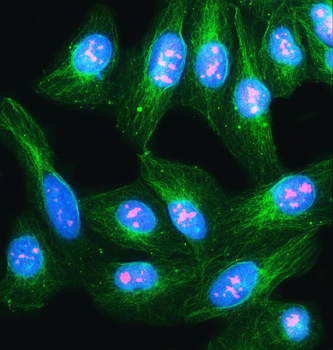
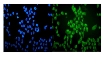
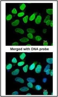
Immunofluorescent analysis of 4% paraformaldehyde-fixed, 0.1% Triton X-100 permeabilized HeLa (human cervical epithelial adenocarcinoma cell line) cells labeling Pdx1 at 1/25 dilution, followed by Dylight 488-conjugated goat anti-mouse IgG secondary antibody at 1/200 dilution (green). Immunofluorescence image showing cytoplasm and nucleus staining on HeLa cell line. Cytoplasmic actin is detected with Dylight 554 Phalloidin at 1/100 dilution (red).
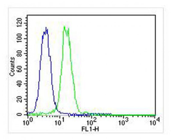
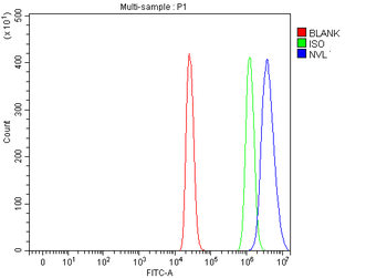
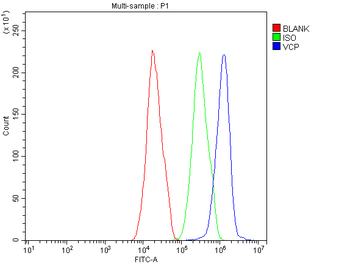
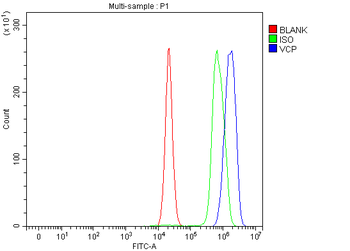
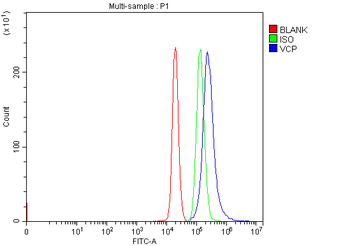
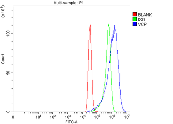
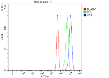
Overlay histogram showing K562 cells stained (green line). The cells were fixed with 2% paraformaldehyde (10 min) and then permeabilized with 90% methanol for 10 min. The cells were then icubated in 2% bovine serum albumin to block non-specific protein-protein interactions followed by the antibody (1:25 dilution) for 60 min at 37°C. The secondary antibody used was Goat-Anti-Mouse IgG, DyLight 488 Conjugated Highly Cross-Adsorbed at 1/400 dilution for 40 min at 37°C. Isotype control antibody (blue line) was mouse IgG1 (1 μg/1x10^6 cells) used under the same conditions. Acquisition of >10000 events was performed.
Quick Database Links
Gene Symbol
VCP
UniProt
UniProt Details
− No UniProt data available
Documents Download
Datasheet
Product Information
Request a Document
Protocol Information
WB
Western Blot (IB, immunoblot)
IHC-P
Immunohistochemistry Paraffin
FC
Flow Cytometry
IF
Immunofluorescence
VCP Antibody (orb1787867)

- 0.0
Based on 0 reviews
Participating in our Biorbyt product reviews program enables you to support fellow scientists by sharing your firsthand experience with our products.
Login to Submit a Review