You have no items in your shopping cart.
Featured
Description
Research Area
Immunology & Inflammation
Images & Validation
−Item 1 of 4
| Tested Applications | IF, IHC-Fr, IHC-P, WB |
|---|---|
| Dilution Range | WB=1:500-2000, IHC-P=1:400-800, IHC-F=1:400-800, IF=1:100-500 |
| Reactivity | Human, Mouse, Rat |
| Predicted Reactivity | Bovine |
Related Conjugates & Formulations
−Key Properties
−| Antibody Type | Primary Antibody |
|---|---|
| Host | Rabbit |
| Clonality | Polyclonal |
| Isotype | IgG |
| Immunogen | KLH conjugated synthetic peptide derived from mouse UMOD (351-450/642aa) |
| Target | UMOD |
| Molecular Weight | 37 kDa |
| Purification | Affinity purified by Protein A |
| Conjugation | Unconjugated |
Storage & Handling
−| Storage | Maintain refrigerated at 2-8°C for up to 2 weeks. For long term storage store at -20°C in small aliquots to prevent freeze-thaw cycles. |
|---|---|
| Form/Appearance | Liquid |
| Buffer/Preservatives | 0.01M TBS (pH7.4) with 1% rAlbumin, 0.02% Proclin300 and 50% Glycerol. |
| Concentration | 1mg/ml |
| Expiration Date | 12 months from date of receipt. |
| Disclaimer | For research use only |
Alternative Names
−ADMCKD2; ADTKD1; FJHN; HNFJ; HNFJ1; MCKD2; THGP; THP; UROM_HUMAN; UMOD; Tamm-Horsfall urinary glycoprotein (THP); uromodulin; uromodulin (uromucoid, Tamm-Horsfall glycoprotein); Tamm-Horsfall glycoprotein; uromucoid
Similar Products
−- Item 1 of 6
Uromucoid Polyclonal Antibody [orb1417658]
ELISA, IF, IHC-Fr, IHC-P, WB
Rat
Rabbit
Polyclonal
Unconjugated
100 μl - Item 1 of 4
UMOD Rabbit Polyclonal Antibody [orb669141]
ELISA, FC, IHC, WB
Human, Mouse, Rat
Rabbit
Polyclonal
Unconjugated
100 μg - Item 1 of 3
UMOD Rabbit Polyclonal Antibody [orb632414]
ELISA, IHC, IP, WB
Human, Mouse, Rat
Rabbit
Polyclonal
Unconjugated
50 μg, 100 μg - Item 1 of 2
THP rabbit pAb Antibody [orb770352]
ELISA, IHC, WB
Human, Mouse, Rat
Polyclonal
Unconjugated
50 μl, 100 μl - Item 1 of 2

Quality Guarantee
Explore bioreagents carefree to elevate your research. All our products are rigorously tested for performance. If a product does not perform as described on its datasheet, our scientific support team will provide expert troubleshooting, a prompt replacement, or a refund. For full details, please see our Terms & Conditions and Buying Guide. Contact us at [email protected].

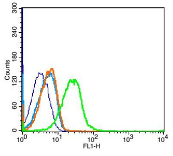
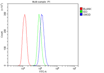
Blank control: Mouse kidney (blue). Primary Antibody: Rabbit Anti-Coxsackie Adenovirus Receptor antibody (orb5637, Green), dilution: 1 µg in 100 µl 1X PBS containing 0.5% BSA, Isotype Control Antibody: Rabbit IgG (orange), used under the same conditions, Secondary Antibody: Goat anti-rabbit IgG-FITC (white blue), dilution: 1:200 in 1X PBS containing 0.5% BSA. Protocol, The cells were fixed with 2% paraformaldehyde for 10 min at 37°C. Primary antibody (orb5637, 1 µg/1x10^6 cells) were incubated for 30 min at room temperature, followed by 1X PBS containing 0.5% BSA + 10% goat serum (1 hour) to block non-specific protein-protein interactions. Then the Goat Anti-rabbit IgG/FITC antibody was added into the blocking buffer mentioned above to react with the primary antibody at 1/200 dilution for 40 min at room temperature. Acquisition of 20000 events was performed.
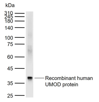
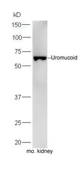
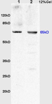
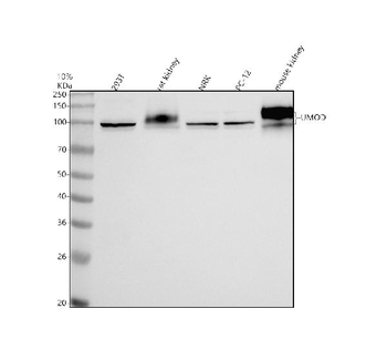
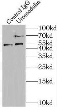
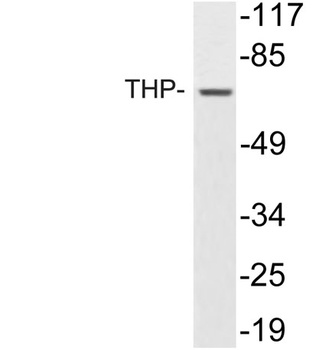
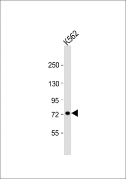
Sample: Lane 1: Recombinant human UMOD protein, N-His, Primary: Anti-UMOD (orb5637) at 1/1000 dilution, Secondary: IRDye800CW Goat Anti-Rabbit IgG at 1/20000 dilution, Predicted band size: 69 kDa, Observed band size: 37 kDa.
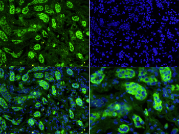
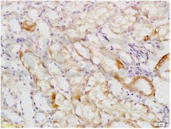
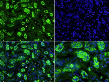
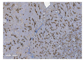
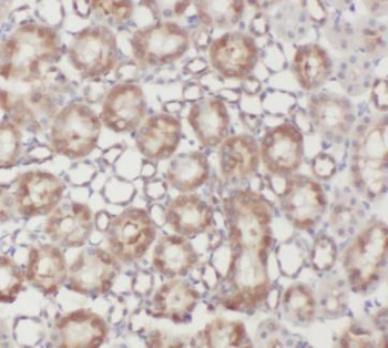
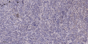
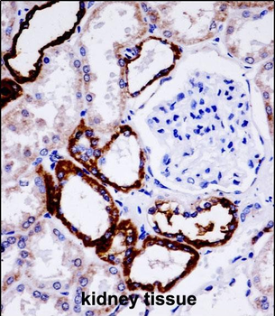
Tissue/Cell: human kidney tissue, 4% Paraformaldehyde-fixed and paraffin-embedded, Antigen retrieval: citrate buffer (0.01M, pH 6.0), Boiling bathing for 15 min, Blocking buffer (normal goat serum) at 37°C for 20 min, Incubation: Anti-Uromucoid Polyclonal Antibody, Unconjugated (orb5637) 1:200, overnight at 4°C, The secondary antibody was Goat Anti-Rabbit IgG, Cy3 conjugated (orb868805) used at 1:200 dilution for 40 minutes at 37°C. DAPI (5 ug/ml, blue) was used to stain the cell nuclei.
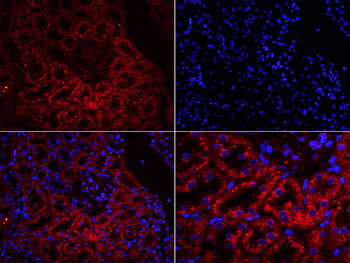
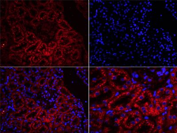
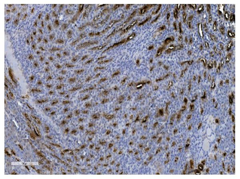
Tissue/Cell: rat kidney tissue, 4% Paraformaldehyde-fixed and paraffin-embedded, Antigen retrieval: citrate buffer (0.01M, pH 6.0), Boiling bathing for 15 min, Blocking buffer (normal goat serum) at 37°C for 20 min, Incubation: Anti-Uromucoid Polyclonal Antibody, Unconjugated (orb5637) 1:200, overnight at 4°C, The secondary antibody was Goat Anti-Rabbit IgG, Cy3 conjugated (orb868589) used at 1:200 dilution for 40 minutes at 37°C. DAPI (5 ug/ml, blue) was used to stain the cell nuclei.
Quick Database Links
Gene Symbol
UMOD
Documents Download
Datasheet
Product Information
Request a Document
Protocol Information
WB
Western Blot (IB, immunoblot)
IHC-P
Immunohistochemistry Paraffin
IHC-Fr
Immunohistochemistry Frozen
IF
Immunofluorescence
UMOD Rabbit Polyclonal Antibody (orb5637)

- 0.0
Based on 0 reviews
Participating in our Biorbyt product reviews program enables you to support fellow scientists by sharing your firsthand experience with our products.
Login to Submit a ReviewAvailable Sizes
Select a size below
Free Secondary Antibody (20 ul)0/0
Please add an antibody product to your cart first.