You have no items in your shopping cart.
Featured
Description
Research Area
Signal Transduction
Images & Validation
−Item 1 of 3
| Tested Applications | AM, IHC, IP, WB |
|---|---|
| Dilution Range | WB (1:1000), IHC (1:1000), ICC/IF (1:100) |
| Reactivity | Human, Mouse, Rat |
| Application Notes |
Key Properties
−| Host | Mouse |
|---|---|
| Clonality | Monoclonal |
| Isotype | IgG2b |
| Clone No. | N67/15 (Formerly sold as S67-15) |
| Immunogen | Synthetic peptide amino acids 827-845 of human TrpC5 (also known as short transient receptor potential channel 5, and Htrp5) |
| Target | TRPC5 |
| Molecular Weight | 110kDa |
| Purification | Protein G Purified |
| Conjugation | Biotin |
Storage & Handling
−| Storage | Conjugated antibodies should be stored according to the product label |
|---|---|
| Buffer/Preservatives | 136.36mM Ethanolamine, 133.23 mM Chlorides, 9.55mM Phosphates, 9.55mM Sodium Bicarbonate |
| Concentration | 1 mg/ml |
| Expiration Date | 12 months from date of receipt. |
| Disclaimer | For research use only |
Alternative Names
−Htrp5, Short transient receptor potential channel 5, TRP5
Similar Products
−- Item 1 of 1
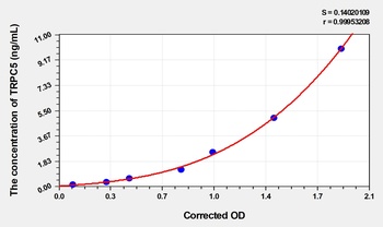
Mouse Transient Receptor Potential Cation Channel Subfamily C Member 5 (TRPC5) ELISA Kit [orb1736785]
Mouse
0.16-10 ng/mL
0.062 ng/mL
48 T, 96 T 
TRPC5 Rabbit Polyclonal Antibody (Biotin) [orb2622841]
ELISA, IHC, WB
Human
Rabbit
Polyclonal
Biotin
100 μgTRPC5 Rabbit Polyclonal Antibody (Biotin) [orb2609479]
ELISA, IHC, WB
Human, Mouse, Rat
Rabbit
Polyclonal
Biotin
100 μg
Quality Guarantee
Explore bioreagents carefree to elevate your research. All our products are rigorously tested for performance. If a product does not perform as described on its datasheet, our scientific support team will provide expert troubleshooting, a prompt replacement, or a refund. For full details, please see our Terms & Conditions and Buying Guide. Contact us at [email protected].

Immunocytochemistry/Immunofluorescence analysis using Mouse Anti-TrpC5 Monoclonal Antibody, Clone N67/15. Tissue: Neuroblastoma cells (SH-SY5Y). Species: Human. Fixation: 4% PFA for 15 min. Primary Antibody: Mouse Anti-TrpC5 Monoclonal Antibody at 1:50 for overnight at 4°C with slow rocking. Secondary Antibody: AlexaFluor 488 at 1:1000 for 1 hour at RT. Counterstain: Phalloidin-iFluor 647 (red) F-Actin stain; Hoechst (blue) nuclear stain at 1:800, 1.6mM for 20 min at RT. (A) Hoechst (blue) nuclear stain. (B) Phalloidin-iFluor 647 (red) F-Actin stain. (C) TrpC5 Antibody (D) Composite.
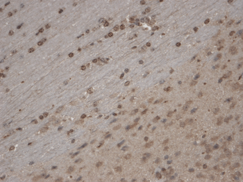
Immunohistochemistry analysis using Mouse Anti-TrpC5 Monoclonal Antibody, Clone N67/15. Tissue: Brain Slice. Species: Mouse. Fixation: 10% Formalin Solution for 12-24 hours at RT. Primary Antibody: Mouse Anti-TrpC5 Monoclonal Antibody at 1:1000 for 1 hour at RT. Secondary Antibody: HRP/DAB Detection System: Biotinylated Goat Anti-Mouse, Streptavidin Peroxidase, DAB Chromogen (brown) for 30 minutes at RT. Counterstain: Mayer Hematoxylin (purple/blue) nuclear stain at 250-500 μl for 5 minutes at RT. Localization: Nuclear staining. Magnification: 10X.
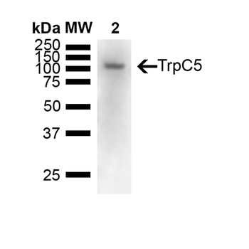
Western Blot analysis of Mouse brain showing detection of 110 kDa TrpC5 protein using Mouse Anti-TrpC5 Monoclonal Antibody, Clone N67/15. Lane 1: Molecular Weight Ladder (MW). Lane 2: Mouse Brain. Load: 15 ug. Block: 5% Skim Milk powder in TBST. Primary Antibody: Mouse Anti-TrpC5 Monoclonal Antibody at 1:1000 for Overnight at 4°C. Secondary Antibody: Goat anti-mouse IgG:HRP at 1:7000 for 1 hour at RT with shaking. Color Development: Chemiluminescent for HRP (Moss) for 5 min in RT. Predicted/Observed Size: 110 kDa.
Quick Database Links
UniProt Details
− No UniProt data available
NCBI Gene Details
− No NCBI Gene data available
NCBI Reference Sequences
−Associated Accession Numbers
Curated reference sequences for the gene transcript and protein product| Protein | NP_036603.1 |
|---|
Documents Download
Datasheet
Product Information
Request a Document
Protocol Information
WB
Western Blot (IB, immunoblot)
IHC
Immunohistochemistry
IP
Immunoprecipitation
TRPC5 Antibody (Biotin) (orb148875)

- 0.0
Based on 0 reviews
Participating in our Biorbyt product reviews program enables you to support fellow scientists by sharing your firsthand experience with our products.
Login to Submit a ReviewAvailable Sizes
Select a size below
Choose Conjugation or Carrier Free Version
Free Secondary Antibody (20 ul)0/0
Please add an antibody product to your cart first.