You have no items in your shopping cart.
Description
Research Area
Signal Transduction
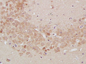
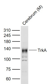
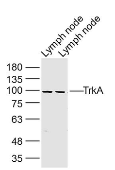
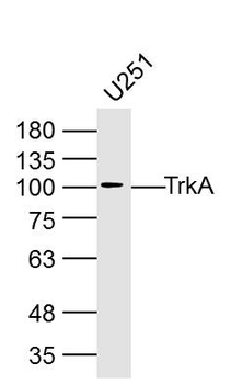
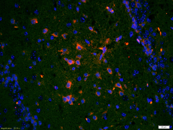
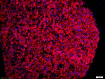
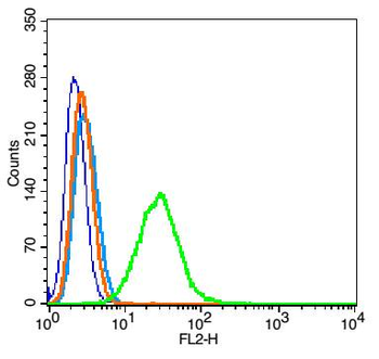
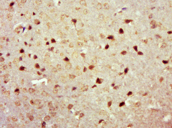
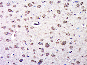
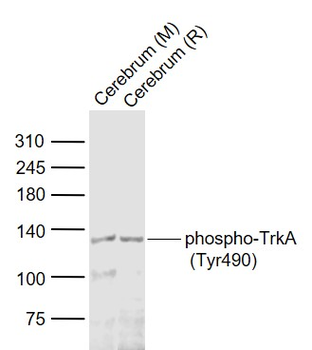
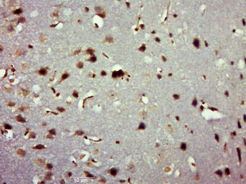
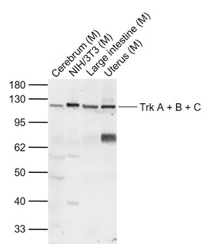
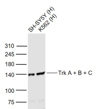
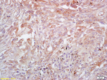
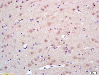
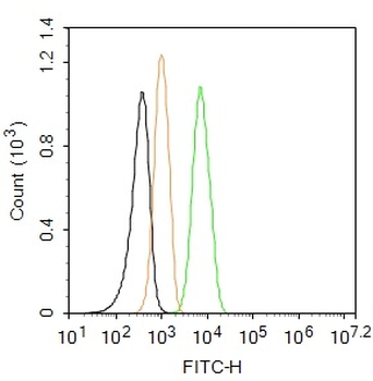
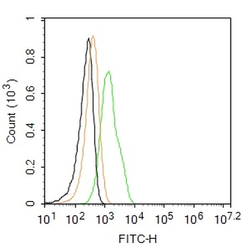
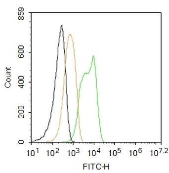
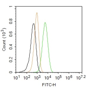
Images & Validation
−| Tested Applications | FC, IF, WB |
|---|---|
| Dilution Range | IF: 1:10~50, WB: 1:1000, FC: 1:10~50 |
| Reactivity | Human, Mouse |
Key Properties
−| Antibody Type | Primary Antibody |
|---|---|
| Host | Rabbit |
| Clonality | Polyclonal |
| Isotype | Rabbit IgG |
| Clone No. | RB17710 |
| Target | This TrkA antibody is generated from rabbits immunized with TrkA his fusion protein |
| Molecular Weight | 87497 Da |
| Conjugation | Unconjugated |
Storage & Handling
−| Storage | Maintain refrigerated at 2-8°C for up to 2 weeks. For long term storage store at -20°C in small aliquots to prevent freeze-thaw cycles |
|---|---|
| Form/Appearance | Purified polyclonal antibody supplied in PBS with 0.09% (W/V) sodium azide. This antibody is prepared by Saturated Ammonium Sulfate (SAS) precipitation followed by dialysis against PBS. |
| Expiration Date | 12 months from date of receipt. |
| Disclaimer | For research use only |
Alternative Names
−High affinity nerve growth factor receptor, Neurotrophic tyrosine kinase receptor type 1, TRK1-transforming tyrosine kinase protein, Tropomyosin-related kinase A, Tyrosine kinase receptor, Tyrosine kinase receptor A, Trk-A, gp140trk, p140-TrkA, NTRK1, MTC, TRK, TRKA
Similar Products
−- Item 1 of 6
TrkA Rabbit Polyclonal Antibody [orb11515]
IF, IHC-Fr, IHC-P, WB
Bovine, Equine, Porcine, Sheep
Human, Mouse, Rat
Rabbit
Polyclonal
Unconjugated
50 μl, 100 μl, 200 μl - Item 1 of 4
Phospho-TrkA (Tyr490) Rabbit Polyclonal Antibody [orb186433]
FC, IF, IHC-Fr, IHC-P, WB
Equine, Gallus, Human, Sheep
Mouse, Rat
Rabbit
Polyclonal
Unconjugated
50 μl, 100 μl, 200 μl - Item 1 of 5
Trk A + B + C Rabbit Polyclonal Antibody [orb11516]
IF, IHC-Fr, IHC-P, WB
Gallus
Human, Mouse, Rat
Rabbit
Polyclonal
Unconjugated
50 μl, 100 μl, 200 μl - Item 1 of 1
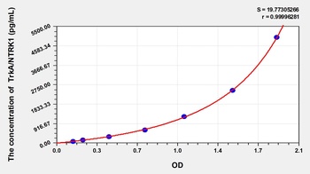
Human High Affinity Nerve Growth Factor Receptor (TrkA/NTRK1) ELISA Kit [orb1146845]
Human
78.13-5000 pg/mL
32 pg/mL
48 T, 96 T - Item 1 of 4
TrkA Rabbit Polyclonal Antibody (FITC) [orb16443]
IF
Bovine, Equine, Porcine, Sheep
Human, Mouse, Rat
Rabbit
Polyclonal
FITC
100 μl
Quality Guarantee
Explore bioreagents carefree to elevate your research. All our products are rigorously tested for performance. If a product does not perform as described on its datasheet, our scientific support team will provide expert troubleshooting, a prompt replacement, or a refund. For full details, please see our Terms & Conditions and Buying Guide. Contact us at [email protected].
Quick Database Links
Gene Symbol
This TrkA antibody is generated from rabbits immunized with TrkA his fusion protein
UniProt
RefSeq (Protein):NP_002520.2, NP_001012331.1, NP_001007793.1
UniProt Details
− No UniProt data available
NCBI Reference Sequences
−Associated Accession Numbers
Curated reference sequences for the gene transcript and protein product| Protein | NP_002520.2, NP_001012331.1, NP_001007793.1 |
|---|
Documents Download
Datasheet
Product Information
Request a Document
Protocol Information
WB
Western Blot (IB, immunoblot)
FC
Flow Cytometry
IF
Immunofluorescence
TrkA Antibody (orb1928981)

- 0.0
Based on 0 reviews
Participating in our Biorbyt product reviews program enables you to support fellow scientists by sharing your firsthand experience with our products.
Login to Submit a ReviewAvailable Sizes
Select a size below
Free Secondary Antibody (20 ul)0/0
Please add an antibody product to your cart first.