You have no items in your shopping cart.
Description
Research Area
Cell Biology
Images & Validation
−Item 1 of 6
| Tested Applications | ICC, IF, IHC, IP, WB |
|---|---|
| Dilution Range | WB (1:1000), IHC (1:100), ICC/IF (1:100) |
| Reactivity | Bovine, Canine, Human, Monkey, Mouse, Rabbit, Rat |
| Application Notes |
Key Properties
−| Host | Rabbit |
|---|---|
| Clonality | Polyclonal |
| Immunogen | Peptide corresponding to AA 20-43 of the mouse TNF-R1 sequence, identical to rat and human over those residues |
| Target | TNF-R1 |
| Molecular Weight | 55kDa |
| Purification | Peptide Affinity Purified |
| Conjugation | PerCP |
Storage & Handling
−| Storage | Conjugated antibodies should be stored according to the product label |
|---|---|
| Buffer/Preservatives | 95.46mM Phosphate, 2.48mM MES and 2mM EDTA |
| Concentration | 1 mg/ml |
| Expiration Date | 12 months from date of receipt. |
| Disclaimer | For research use only |
Alternative Names
−Tumor necrosis factor receptor 1, TNFR-1, TNFRSF1A, TNFAR, TNFR1
Similar Products
−
CD120a/TNFRSF1A Mouse Monoclonal Antibody (PerCP) [orb2971948]
FC
Human
Mouse
Monoclonal
PerCP
50 T, 100 TTNFR1 Rabbit Polyclonal Antibody (PerCP-Cy7) [orb1591936]
FC
Bovine, Canine, Equine, Porcine, Rabbit
Human, Mouse, Rat
Rabbit
Polyclonal
PerCP/Cy7
100 μlTNFR1 Rabbit Polyclonal Antibody (PerCP-Cy5.5) [orb1591937]
FC
Bovine, Canine, Equine, Porcine, Rabbit
Human, Mouse, Rat
Rabbit
Polyclonal
PerCP/Cy5.5
100 μlTNFR1 Rabbit Polyclonal Antibody (PerCP) [orb1591938]
FC
Bovine, Canine, Equine, Porcine, Rabbit
Human, Mouse, Rat
Rabbit
Polyclonal
PerCP
100 μlTNFR1 Rabbit Polyclonal Antibody (PerCP-Cy5.5) [orb2449714]
IF
Human
Rabbit
Polyclonal
PerCP/Cy5.5
100 μl
Quality Guarantee
Explore bioreagents carefree to elevate your research. All our products are rigorously tested for performance. If a product does not perform as described on its datasheet, our scientific support team will provide expert troubleshooting, a prompt replacement, or a refund. For full details, please see our Terms & Conditions and Buying Guide. Contact us at [email protected].
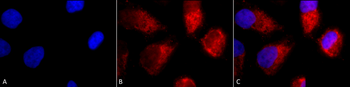
Immunocytochemistry/Immunofluorescence analysis using Rabbit Anti-TNF-R1 Polyclonal Antibody. Tissue: Cervical cancer cell line (HeLa). Species: Human. Fixation: 2% Formaldehyde for 20 min at RT. Primary Antibody: Rabbit Anti-TNF-R1 Polyclonal Antibody at 1:100 for 12 hours at 4°C. Secondary Antibody: APC Goat Anti-Rabbit (red) at 1:200 for 2 hours at RT. Counterstain: DAPI (blue) nuclear stain at 1:40000 for 2 hours at RT. Localization: Golgi apparatus membrane. Magnification: 100x. (A) DAPI (blue) nuclear stain. (B) Anti-TNF-R1 Antibody. (C) Composite.
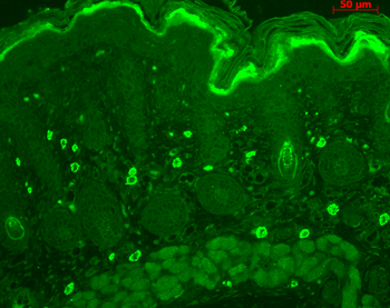
Immunohistochemistry analysis using Rabbit Anti-TNF-R1 Polyclonal Antibody. Tissue: backskin. Species: Mouse. Fixation: Bouin's Fixative Solution. Primary Antibody: Rabbit Anti-TNF-R1 Polyclonal Antibody at 1:100 for 1 hour at RT. Secondary Antibody: FITC Goat Anti-Rabbit (green) at 1:50 for 1 hour at RT. Localization: dermis.
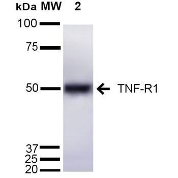
Western blot analysis of Mouse Liver cell lysates showing detection of ~55 kDa TNF-R1 protein using Rabbit Anti-TNF-R1 Polyclonal Antibody. Lane 1: Molecular Weight Ladder (MW). Lane 2: Mouse Liver cell lysates. Load: 15 μg. Block: 5% Skim Milk in 1X TBST. Primary Antibody: Rabbit Anti-TNF-R1 Polyclonal Antibody at 1:1000 for 2 hours at RT. Secondary Antibody: Goat Anti-Rabbit IgG: HRP at 1:2000 for 60 min at RT. Color Development: ECL solution for 5 min at RT. Predicted/Observed Size: ~55 kDa.
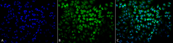
Immunocytochemistry/Immunofluorescence analysis using Rabbit Anti-TNF-R1 Polyclonal Antibody. Tissue: Cervical cancer cell line (HeLa). Species: Human. Fixation: 2% Formaldehyde for 20 min at RT. Primary Antibody: Rabbit Anti-TNF-R1 Polyclonal Antibody at 1:100 for 12 hours at 4°C. Secondary Antibody: FITC Goat Anti-Rabbit (green) at 1:200 for 2 hours at RT. Counterstain: DAPI (blue) nuclear stain at 1:40000 for 2 hours at RT. Localization: Golgi apparatus membrane. Magnification: 20x. (A) DAPI (blue) nuclear stain. (B) Anti-TNF-R1 Antibody. (C) Composite.
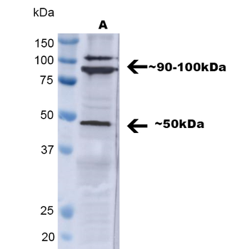
Western blot analysis of Human A549 showing detection of ~ 50 kDa TNF-R1 protein using Rabbit Anti-TNF-R1 Polyclonal Antibody. Lane 1: MW Ladder, Lane 2: A549. Load: 30 ug. Block: 5% BSA in TBST. Primary Antibody: Rabbit Anti-TNF-R1 Polyclonal Antibody at 1:1000 for 2 hours at RT with shaking. Secondary Antibody: Goat Anti-Rabbit IgG: HRP at 1:4000 for 1 hour at RT with shaking. Color Development: Chemiluminescent for HRP (Moss) for 5 min in RT. Predicted/Observed Size: ~ 50 kDa. Other Band (s): ~90-100kDa. Other bands can be explained by a few factors, such as oligomerization, self-aggregation, cleavage of the TNFR1 extracellular domain, etc.
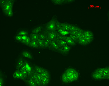
Immunocytochemistry/Immunofluorescence analysis using Rabbit Anti-TNF-R1 Polyclonal Antibody. Tissue: HaCaT cells. Species: Human. Fixation: Cold 100% methanol at -20°C for 10 minutes. Primary Antibody: Rabbit Anti-TNF-R1 Polyclonal Antibody at 1:100 for 12 hours at 4°C. Secondary Antibody: FITC Goat Anti-Rabbit at 1:50 for 1-2 hours at RT in dark. Localization: Punctate nuclear staining, dotty staining in cytoplasm.
UniProt Details
− No UniProt data available
NCBI Gene Details
− No NCBI Gene data available
NCBI Reference Sequences
−Associated Accession Numbers
Curated reference sequences for the gene transcript and protein product| RefSeq | P19438 |
|---|
Documents Download
Datasheet
Product Information
Request a Document
Protocol Information
WB
Western Blot (IB, immunoblot)
IHC
Immunohistochemistry
IF
Immunofluorescence
ICC
Immunocytochemistry
IP
Immunoprecipitation
TNF-R1 Antibody (PerCP) (orb151866)

- 0.0
Based on 0 reviews
Participating in our Biorbyt product reviews program enables you to support fellow scientists by sharing your firsthand experience with our products.
Login to Submit a ReviewAvailable Sizes
Select a size below
Choose Conjugation or Carrier Free Version
Free Secondary Antibody (20 ul)0/0
Please add an antibody product to your cart first.