You have no items in your shopping cart.
Featured
Description
Research Area
Metabolism Research
Images & Validation
−Item 1 of 4
| Tested Applications | ELISA, IHC, WB |
|---|---|
| Dilution Range | WB (1:500); IHC (1:100) |
| Reactivity | Human, Mouse, Rat |
| Application Notes |
Key Properties
−| Host | Mouse |
|---|---|
| Clonality | Monoclonal |
| Isotype | IgG2a |
| Clone No. | H43 |
| Immunogen | Synthetic peptide from the full length Human Thyroid hormone receptor protein |
| Target | Thyroid Hormone Receptor |
| Molecular Weight | 70 kDa |
| Purification | Protein A Purified |
| Conjugation | Biotin |
Storage & Handling
−| Storage | Conjugated antibodies should be stored according to the product label |
|---|---|
| Buffer/Preservatives | 136.36mM Ethanolamine, 133.23 mM Chlorides, 9.55mM Phosphates, 9.55mM Sodium Bicarbonate |
| Concentration | 1mg/ml |
| Expiration Date | 12 months from date of receipt. |
| Disclaimer | For research use only |
Alternative Names
−ERBA ALPHA, ERBA beta, ERBA1, ERBA2, NR1A1, NR1A2, THR1, THRA, THRA1, THRA2, THRB, THRB1, THRB2, Thyroid hormone receptor alpha, Thyroid hormone receptor beta
Similar Products
−- Item 1 of 1
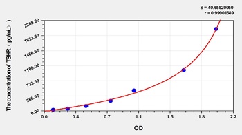
Goat Thyroid Stimulating Hormone Receptor (TSHR) ELISA Kit [orb1146796]
Goat
31.25-2000 pg/mL
10.8 pg/mL
48 T, 96 T - Item 1 of 1
Human Anti-Thyroid Stimulating Hormone Receptor Antiboby (TSHRAb) ELISA Kit [orb1146792]
Human
1.57-100 mIU/mL
0.82 mIU/mL
48 T, 96 T - Item 1 of 1
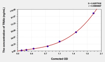
Mouse Thyroid Stimulating Hormone Receptor Antibody (TRAb) ELISA Kit [orb1817325]
Mouse
1.57-100 ng/mL
0.63 ng/mL
48 T, 96 T 

Quality Guarantee
Explore bioreagents carefree to elevate your research. All our products are rigorously tested for performance. If a product does not perform as described on its datasheet, our scientific support team will provide expert troubleshooting, a prompt replacement, or a refund. For full details, please see our Terms & Conditions and Buying Guide. Contact us at [email protected].
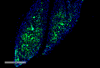
Immunohistochemistry analysis using Mouse Anti-Thyroid Hormone Receptor Monoclonal Antibody, Clone H43. Tissue: Thyroid. Species: Mouse. Primary Antibody: Mouse Anti-Thyroid Hormone Receptor Monoclonal Antibody at 1:100 for Overnight at 4°C, then 30 min at 37°C. Secondary Antibody: Goat Anti-Mouse IgG (H+L): FITC for 45 min at 37°C. Counterstain: DAPI for 3 min at RT. Magnification: 7.5X.
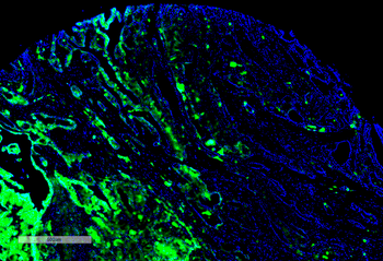
Immunohistochemistry analysis using Mouse Anti-Thyroid Hormone Receptor Monoclonal Antibody, Clone H43. Tissue: Thyroid Cancer. Species: Human. Primary Antibody: Mouse Anti-Thyroid Hormone Receptor Monoclonal Antibody at 1:100 for Overnight at 4°C, then 30 min at 37°C. Secondary Antibody: Goat Anti-Mouse IgG (H+L): FITC for 45 min at 37°C. Counterstain: DAPI for 3 min at RT. Magnification: 4X.
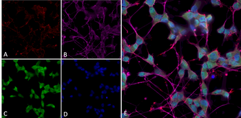
Immunocytochemistry/Immunofluorescence analysis using Mouse Anti-Thyroid Hormone Receptor Monoclonal Antibody, Clone H43. Tissue: Differentiated SH-SY5Y. Species: Human. Primary Antibody: Mouse Anti-Thyroid Hormone Receptor Monoclonal Antibody at 1:250. Secondary Antibody: AlexaFluor 488. Counterstain: phalloidin (Alexa 647, red), beta tubulin (Anti-beta III Tubulin Ab, Alexa 555, magenta) Hoechst (blue). (A) Phalloidin (B) Anti-beta III Tubulin Ab. (C) Thyroid Hormone Receptor Antibody. (D) Hoechst (E) Composite.
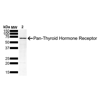
Western Blot analysis of Human Hep G2 Hepatoblastoma Cell lysate showing detection of Thyroid Hormone Receptor protein using Mouse Anti-Thyroid Hormone Receptor Monoclonal Antibody, Clone H43. Load: 10 ug. Primary Antibody: Mouse Anti-Thyroid Hormone Receptor Monoclonal Antibody at 1:500 for 2 hours at RT with shaking. Secondary Antibody: Goat anti-mouse IgG:HRP at 1:4000 for 1 hour at RT with shaking. Color Development: Chemiluminescent for HRP (Moss) for 5 min in RT. Other Band (s): Higher molceular weight bands could be due to PTMs.
Quick Database Links
UniProt Details
− No UniProt data available
NCBI Gene Details
− No NCBI Gene data available
NCBI Reference Sequences
−Associated Accession Numbers
Curated reference sequences for the gene transcript and protein product| mRNA | NM_001190918.2 |
|---|
Documents Download
Datasheet
Product Information
Request a Document
Protocol Information
WB
Western Blot (IB, immunoblot)
IHC
Immunohistochemistry
ELISA
Enzyme-linked Immunosorbent Assay (EIA)
Thyroid Hormone Receptor Antibody (Biotin) (orb612668)

- 0.0
Based on 0 reviews
Participating in our Biorbyt product reviews program enables you to support fellow scientists by sharing your firsthand experience with our products.
Login to Submit a ReviewAvailable Sizes
Select a size below
Choose Conjugation or Carrier Free Version
Free Secondary Antibody (20 ul)0/0
Please add an antibody product to your cart first.