
You have no items in your shopping cart.
Description
Research Area
Epigenetics
Images & Validation
−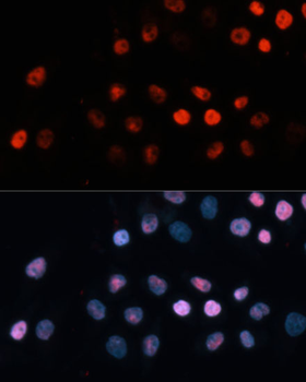
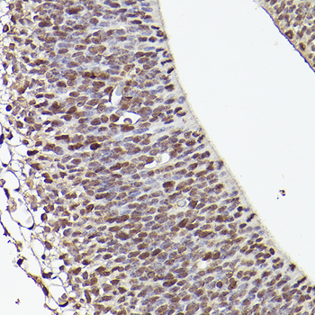
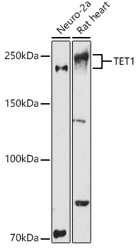
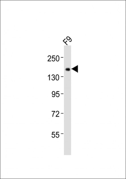
Item 1 of 3
| Tested Applications | ELISA, IF, IHC-P, WB |
|---|---|
| Dilution Range | WB: 1:500-1:2000, IP: 1:20-1:100 |
| Reactivity | Human, Mouse, Rat |
Key Properties
−| Host | Rabbit |
|---|---|
| Clonality | Polyclonal |
| Isotype | IgG |
| Immunogen | A synthetic peptide corresponding to a sequence within amino acids 1800-1900 of human TET1 (NP_085128.2). |
| Molecular Weight | 250kDa |
| Purification | Affinity purification |
| Conjugation | Unconjugated |
Storage & Handling
−| Storage | Maintain refrigerated at 2-8°C for up to 2 weeks. For long term storage store at -20°C in small aliquots to prevent freeze-thaw cycles. |
|---|---|
| Buffer/Preservatives | PBS with 0.05% proclin300,50% glycerol,pH7.3. |
| Expiration Date | 12 months from date of receipt. |
| Disclaimer | For research use only |
Alternative Names
−TET1, CXXC6, LCX, bA119F7.1, MLL-TET1, TET1-MLL
Similar Products
−- Item 1 of 1

TET1 Rabbit pAb, Pacific Blue conjugated [orb2864416]
FC, ICC, IF
Bovine, Canine, Equine, Gallus, Porcine, Rabbit, Rat
Human, Mouse
Rabbit
Polyclonal
Pacific Blue
100 μl

Quality Guarantee
Explore bioreagents carefree to elevate your research. All our products are rigorously tested for performance. If a product does not perform as described on its datasheet, our scientific support team will provide expert troubleshooting, a prompt replacement, or a refund. For full details, please see our Terms & Conditions and Buying Guide. Contact us at [email protected].
Quick Database Links
UniProt
UniProt Details
− No UniProt data available
Protocol Information
WB
Western Blot (IB, immunoblot)
IHC-P
Immunohistochemistry Paraffin
IF
Immunofluorescence
ELISA
Enzyme-linked Immunosorbent Assay (EIA)
Filter by Applications
Filter by Species
Xiaojun Zhou 1 2, Shan Jiang 3 4, Siyi Guo 4, Shuai Yao 4, Qiqi Sheng 4, Qian Zhang 5, Jianjun Dong 4, Lin Liao C/EBPβ-Lin28a positive feedback loop triggered by C/EBPβ hypomethylation enhances the proliferation and migration of vascular smooth muscle cells in restenosis Chin Med J (Engl), (2024)
Applications
WB
Reactivity
Rat