You have no items in your shopping cart.
Featured
Description
Research Area
Cancer Biology, Cell Biology, Protein Biochemistry, Signal Transduction
Images & Validation
−Item 1 of 3
| Tested Applications | FC, ICC, IF, IP, WB |
|---|---|
| Dilution Range | WB (1:1000), ICC/IF (1:100) |
| Reactivity | Bovine, Canine, Drosophila, Guinea pig, Hamster, Human, Monkey, Mouse, Other, Porcine, Rabbit, Rat, Saccharomyces, Yeast |
| Application Notes |
Key Properties
−| Host | Rat |
|---|---|
| Clonality | Monoclonal |
| Isotype | IgG2a |
| Clone No. | 91a |
| Immunogen | Recombinant Mouse TCP1 alpha protein fragment (carboxy terminal region). |
| Target | TCP1 alpha |
| Molecular Weight | 60kDa |
| Purification | Protein G Purified |
| Conjugation | Unconjugated |
Storage & Handling
−| Storage | Maintain refrigerated at 2-8°C for up to 2 weeks. For long term storage store at -20°C in small aliquots to prevent freeze-thaw cycles. |
|---|---|
| Buffer/Preservatives | PBS pH 7.4, 50% glycerol, 0.1% sodium azide. Storage buffer changes when conjugated. |
| Concentration | 1 mg/ml |
| Expiration Date | 12 months from date of receipt. |
| Disclaimer | For research use only |
Alternative Names
−p63, Tcp-1, TCP1 alpha, Ccta, TRic, Tp63, 21454, c-cpn, CCT, Tcp1, ccpn, AI528772, Cct1, Tcp 1, c cpn
Similar Products
−- Item 1 of 7
TCP1 alpha Rabbit Polyclonal Antibody [orb334558]
ICC, IF, IHC, WB
Human, Mouse, Rat
Rabbit
Polyclonal
Unconjugated
100 μg - Item 1 of 4
- Item 1 of 5
TCP1 alpha Mouse Monoclonal Antibody [orb421123]
FC, ICC, IF, IHC, WB
Human
Mouse
Monoclonal
Unconjugated
100 μg - Item 1 of 3


Quality Guarantee
Explore bioreagents carefree to elevate your research. All our products are rigorously tested for performance. If a product does not perform as described on its datasheet, our scientific support team will provide expert troubleshooting, a prompt replacement, or a refund. For full details, please see our Terms & Conditions and Buying Guide. Contact us at [email protected].
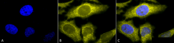
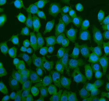
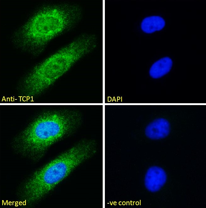
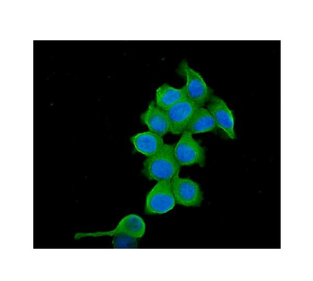
Immunocytochemistry/Immunofluorescence analysis using Rat Anti-TCP1-alpha Monoclonal Antibody, Clone 91a. Tissue: Heat Shocked cervical cancer cells (HeLa). Species: Human. Fixation: 2% Formaldehyde for 20 min at RT. Primary Antibody: Rat Anti-TCP1-alpha Monoclonal Antibody at 1:100 for 12 hours at 4°C. Secondary Antibody: R-PE Goat Anti-Rat (yellow) at 1:200 for 2 hours at RT. Counterstain: DAPI (blue) nuclear stain at 1:40000 for 2 hours at RT. Localization: Cytoplasm. Centrosome. Magnification: 100x. (A) DAPI (blue) nuclear stain. (B) Anti-TCP1-alpha Antibody. (C) Composite. Heat Shocked at 42°C for 1h.
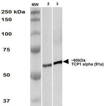
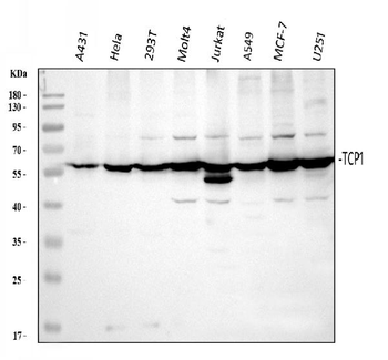
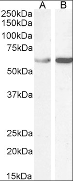
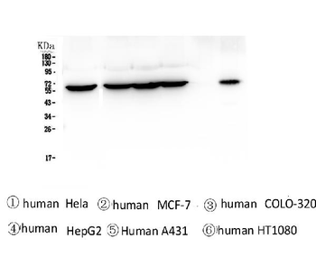
Western Blot analysis of Human A431 and HEK293 cell lysates showing detection of TCP1 alpha protein using Rat Anti-TCP1 alpha Monoclonal Antibody, Clone 91a. Primary Antibody: Rat Anti-TCP1 alpha Monoclonal Antibody at 1:1000.
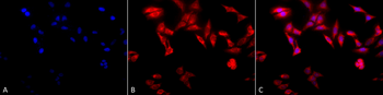
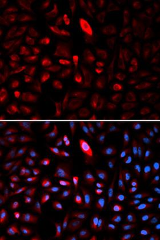
Immunocytochemistry/Immunofluorescence analysis using Rat Anti-TCP1-alpha Monoclonal Antibody, Clone 91a. Tissue: Heat Shocked cervical cancer cells (HeLa). Species: Human. Fixation: 2% Formaldehyde for 20 min at RT. Primary Antibody: Rat Anti-TCP1-alpha Monoclonal Antibody at 1:100 for 12 hours at 4°C. Secondary Antibody: APC Goat Anti-Rat (red) at 1:200 for 2 hours at RT. Counterstain: DAPI (blue) nuclear stain at 1:40000 for 2 hours at RT. Localization: Cytoplasm. Centrosome. Magnification: 20x. (A) DAPI (blue) nuclear stain. (B) Anti-TCP1-alpha Antibody. (C) Composite. Heat Shocked at 42°C for 1h.
Quick Database Links
UniProt Details
− No UniProt data available
NCBI Gene Details
− No NCBI Gene data available
NCBI Reference Sequences
−Associated Accession Numbers
Curated reference sequences for the gene transcript and protein product| Protein | NP_038714.2 |
|---|
Documents Download
Datasheet
Product Information
Request a Document
Protocol Information
WB
Western Blot (IB, immunoblot)
FC
Flow Cytometry
IF
Immunofluorescence
ICC
Immunocytochemistry
IP
Immunoprecipitation
TCP1 alpha Antibody (orb151162)

- 0.0
Based on 0 reviews
Participating in our Biorbyt product reviews program enables you to support fellow scientists by sharing your firsthand experience with our products.
Login to Submit a ReviewAvailable Sizes
Select a size below
Choose Conjugation or Carrier Free Version
Free Secondary Antibody (20 ul)0/0
Please add an antibody product to your cart first.