You have no items in your shopping cart.
Description
Research Area
Cell Biology, Protein Biochemistry, Signal Transduction
Images & Validation
−
Item 1 of 5
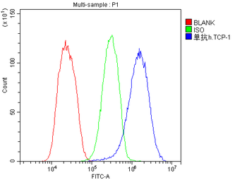
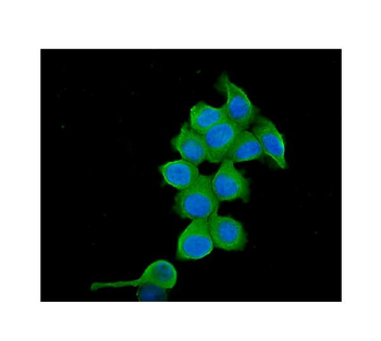
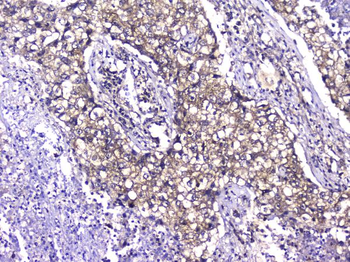
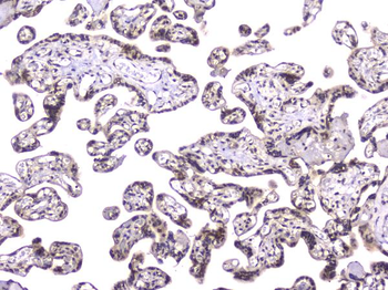
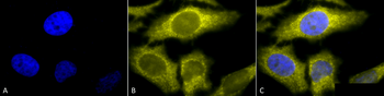
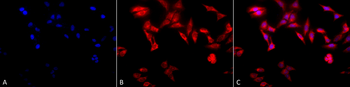
| Tested Applications | FC, ICC, IF, IHC, WB |
|---|---|
| Dilution Range | Western blot, 0.1-0.5μg/ml Immunohistochemistry (Paraffin-embedded Section), 0.5-1μg/ml Immunocytochemistry/Immunofluorescence, 2μg/ml Flow Cytometry (Fixed), 1-3μg/1x10^6 cells |
| Reactivity | Human |
Related Conjugates & Formulations
−Key Properties
−| Antibody Type | Primary Antibody |
|---|---|
| Host | Mouse |
| Clonality | Monoclonal |
| Isotype | Mouse IgG1 |
| Clone No. | B4G9 |
| Immunogen | A synthetic peptide corresponding to a sequence at the C-terminus of human TCP1 alpha, different from the related mouse sequence by one amino acid, and from the related rat sequence by two amino acids. |
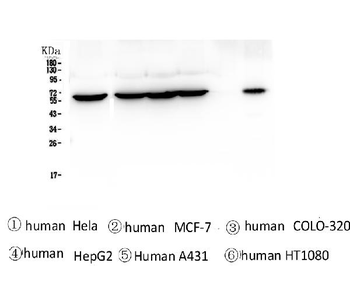
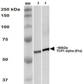
| Target | T-complex protein 1 subunit alpha |
| Molecular Weight | 60 kDa |
| Purification | Immunogen affinity purified. |
| Conjugation | Unconjugated |
Storage & Handling
−| Storage | Maintain refrigerated at 2-8°C for up to 2 weeks. For long term storage store at -20°C in small aliquots to prevent freeze-thaw cycles. |
|---|---|
| Form/Appearance | Lyophilized |
| Buffer/Preservatives | Each vial contains 4mg Trehalose, 0.9mg NaCl, 0.2mg Na2HPO4, 0.05mg NaN3. |
| Concentration | Adding 0.2 ml of distilled water will yield a concentration of 500 μg/ml. |
| Expiration Date | 12 months from date of receipt. |
| Disclaimer | For research use only |
Alternative Names
−CCT alpha; CCT1; CCTa; D6S230E; t complex 1; TCP 1 alpha; TCP1
Similar Products
−- Item 1 of 3
TCP1 alpha Antibody [orb151162]
FC, ICC, IF, IP, WB
Bovine, Canine, Drosophila, Guinea pig, Hamster, Human, Monkey, Mouse, Other, Porcine, Rabbit, Rat, Saccharomyces, Yeast
Rat
Monoclonal
Unconjugated
100 μg 
Quality Guarantee
Explore bioreagents carefree to elevate your research. All our products are rigorously tested for performance. If a product does not perform as described on its datasheet, our scientific support team will provide expert troubleshooting, a prompt replacement, or a refund. For full details, please see our Terms & Conditions and Buying Guide. Contact us at [email protected].
Quick Database Links
Gene Symbol
T-complex protein 1 subunit alpha
UniProt
UniProt Details
− No UniProt data available
Protocol Information
WB
Western Blot (IB, immunoblot)
IHC
Immunohistochemistry
FC
Flow Cytometry
IF
Immunofluorescence
ICC
Immunocytochemistry
Available Sizes
Select a size below
Free Secondary Antibody (20 ul)0/0
Please add an antibody product to your cart first.