You have no items in your shopping cart.
Featured
Description
Research Area
Actin Binding Proteins, Cell Biology, Nuclear envelope
Images & Validation
−Item 1 of 6
| Tested Applications | ICC, IF, IHC-Fr, IHC-P, WB |
|---|---|
| Dilution Range | WB=1:500-2000, IHC-P=1:50-200, IHC-F=1:50-200, ICC/IF=1:50-200, IF=1:50-200 |
| Reactivity | Human, Mouse, Rat |
Key Properties
−| Antibody Type | Primary Antibody |
|---|---|
| Host | Rabbit |
| Clonality | Recombinant |
| Isotype | IgG |
| Clone No. | B33C0 |
| Immunogen | Recombinant protein within human SUN2 aa 500-700. |
| Target | SUN2 |
| Molecular Weight | 80 kDa |
| Purification | Affinity purified by Protein A |
| Conjugation | Unconjugated |
Storage & Handling
−| Storage | Maintain refrigerated at 2-8°C for up to 2 weeks. For long term storage store at -20°C in small aliquots to prevent freeze-thaw cycles. |
|---|---|
| Form/Appearance | Liquid |
| Buffer/Preservatives | 0.01M TBS (pH7.4) with 1% rAlbumin, 0.02% Proclin300 and 50% Glycerol. |
| Concentration | 1mg/ml |
| Expiration Date | 12 months from date of receipt. |
| Disclaimer | For research use only |
Alternative Names
−UNC84B; rab5IP; B230369L08Rik; C030011B15; RGD1563141; SUN2_HUMAN; SUN2; Protein unc-84 homolog B; Rab5-interacting protein (Rab5IP); Sad1/unc-84 protein-like 2; FRIGG; KIAA0668; SUN2_MOUSE; Sad1 and UNC84 domain containing 2; unc-84 homolog B (C. elegans)
Similar Products
−
SUN2 Rabbit Monoclonal Antibody [orb2989564]
WB
Human
Rabbit
Monoclonal
Unconjugated
200 μl, 100 μl, 50 μl, 30 μl
Quality Guarantee
Explore bioreagents carefree to elevate your research. All our products are rigorously tested for performance. If a product does not perform as described on its datasheet, our scientific support team will provide expert troubleshooting, a prompt replacement, or a refund. For full details, please see our Terms & Conditions and Buying Guide. Contact us at [email protected].
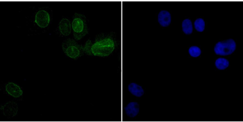
ICC staining SUN2 (green) in SK-Br-3 cells. The nuclear counter stain is DAPI (blue). Cells were fixed in paraformaldehyde, permeabilised with 0.25% Triton X100/PBS.
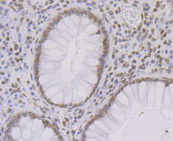
Immunohistochemical analysis of paraffin-embedded human colon tissue using anti-SUN2 antibody. Counter stained with hematoxylin.
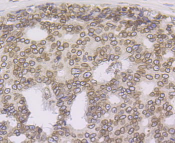
Immunohistochemical analysis of paraffin-embedded human prostate cancer tissue using anti-SUN2 antibody. Counter stained with hematoxylin.

Immunohistochemical analysis of paraffin-embedded mouse liver tissue using anti-SUN2 antibody. Counter stained with hematoxylin.
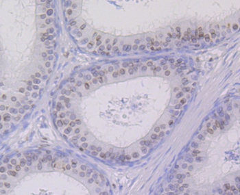
Immunohistochemical analysis of paraffin-embedded rat epididymis tissue using anti-SUN2 antibody. Counter stained with hematoxylin.
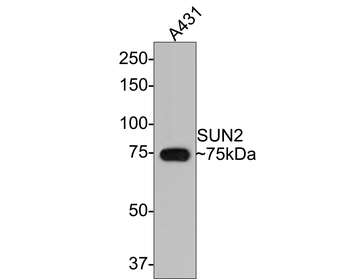
Western blot analysis of SUN2 on A431 cell lysates with Rabbit anti-SUN2 antibody (orb1499408) at 1/500 dilution. Lysates/proteins at 10 µg/Lane. Predicted band size: 80 kDa, Observed band size: 75 kDa, Exposure time: 2 minutes, 8% SDS-PAGE gel. Proteins were transferred to a PVDF membrane and blocked with 5% NFDM/TBST for 1 hour at room temperature. The primary antibody (orb1499408) at 1/500 dilution was used in 5% NFDM/TBST at room temperature for 2 hours. Goat Anti-Rabbit IgG - HRP Secondary Antibody at 1:300000 dilution was used for 1 hour at room temperature.
Quick Database Links
Gene Symbol
SUN2
Documents Download
Datasheet
Product Information
Request a Document
Protocol Information
WB
Western Blot (IB, immunoblot)
IHC-P
Immunohistochemistry Paraffin
IHC-Fr
Immunohistochemistry Frozen
IF
Immunofluorescence
ICC
Immunocytochemistry
SUN2 Recombinant Rabbit Monoclonal Antibody (orb1499408)

- 0.0
Based on 0 reviews
Participating in our Biorbyt product reviews program enables you to support fellow scientists by sharing your firsthand experience with our products.
Login to Submit a ReviewAvailable Sizes
Select a size below
Free Secondary Antibody (20 ul)0/0
Please add an antibody product to your cart first.