You have no items in your shopping cart.
Featured
KO/KD
Validated
Validated
Description
Research Area
Cell Biology
Images & Validation
−Item 1 of 10
| Tested Applications | ELISA, IF, IHC-P, KO/KD Validated, WB |
|---|---|
| Reactivity | Human, Mouse, Rat |
| Application Notes |
Key Properties
−| Antibody Type | Primary Antibody |
|---|---|
| Host | Rabbit |
| Clonality | Polyclonal |
| Isotype | IgG |
| Immunogen | SQSTM1 antibody was raised against a 13 amino acid synthetic peptide from near the carboxy terminus of human SQSTM1.The immunogen is located within the last 50 amino acids of SQSTM1. |
| Target | SQSTM1 |
| Molecular Weight | Predicted: 48kDObserved: 65 kD |
| Purification | SQSTM1 Antibody is affinity chromatography purified via peptide column. |
| Conjugation | Unconjugated |
Storage & Handling
−| Storage | Maintain refrigerated at 2-8°C for up to 2 weeks. For long term storage store at -20°C in small aliquots to prevent freeze-thaw cycles. |
|---|---|
| Form/Appearance | Liquid |
| Buffer/Preservatives | SQSTM1 Antibody is supplied in PBS containing 0.02% sodium azide. |
| Concentration | 1 mg/mL |
| Expiration Date | 12 months from date of receipt. |
| Disclaimer | For research use only |
Alternative Names
−SQSTM1 Antibody: p60, p62, A170, OSIL, PDB3, ZIP3, p62B, ORCA, Sequestosome-1, EBI3-associated protein of 60 kDa, EBIAP
Similar Products
−- Item 1 of 11
SQSTM1 Rabbit Polyclonal Antibody [orb1294360]
IF, IHC, WB
Human, Mouse, Rat
Rabbit
Polyclonal
Unconjugated
100 μl, 25 μl - Item 1 of 9
SQSTM1/p62 Mouse Monoclonal Antibody [orb570318]
FC, ICC, IF, IHC, WB
Human, Mouse, Rat
Mouse
Monoclonal
Unconjugated
100 μg - Item 1 of 8
SQSTM1/p62 Rabbit Polyclonal Antibody [orb507551]
ICC, IF, IHC, WB
Human, Mouse, Rat
Rabbit
Polyclonal
Unconjugated
100 μg - Item 1 of 8
SQSTM1/p62 Recombinant Rabbit Monoclonal Antibody [orb1499273]
FC, ICC, IF, IHC-Fr, IHC-P, WB
Mouse, Rat
Human, Mouse, Rat
Rabbit
Recombinant
Unconjugated
50 μl, 100 μl, 25 μl - Item 1 of 5
SQSTM1/P62 Rabbit Polyclonal Antibody [orb100344]
ICC, IF, IHC-Fr, IHC-P
Bovine, Canine, Equine, Porcine, Rabbit, Sheep
Human, Mouse, Rat
Rabbit
Polyclonal
Unconjugated
50 μl, 100 μl, 200 μl
Quality Guarantee
Explore bioreagents carefree to elevate your research. All our products are rigorously tested for performance. If a product does not perform as described on its datasheet, our scientific support team will provide expert troubleshooting, a prompt replacement, or a refund. For full details, please see our Terms & Conditions and Buying Guide. Contact us at [email protected].
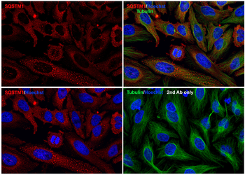
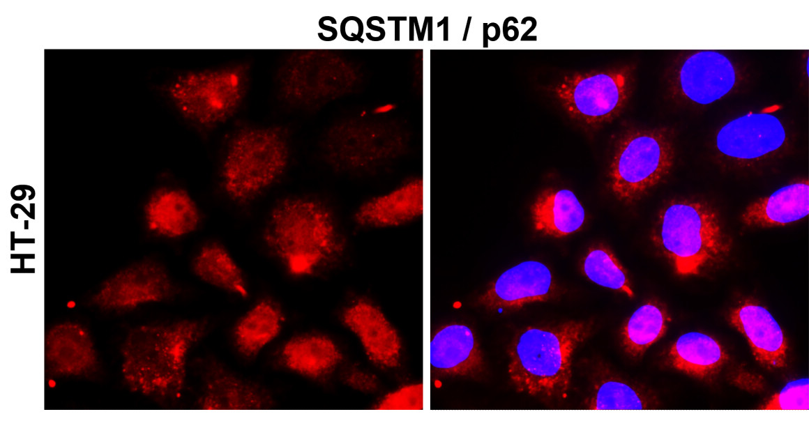
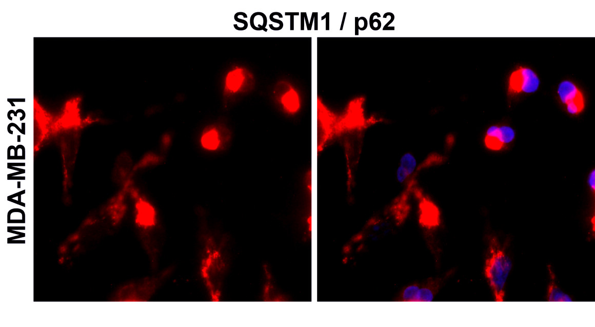
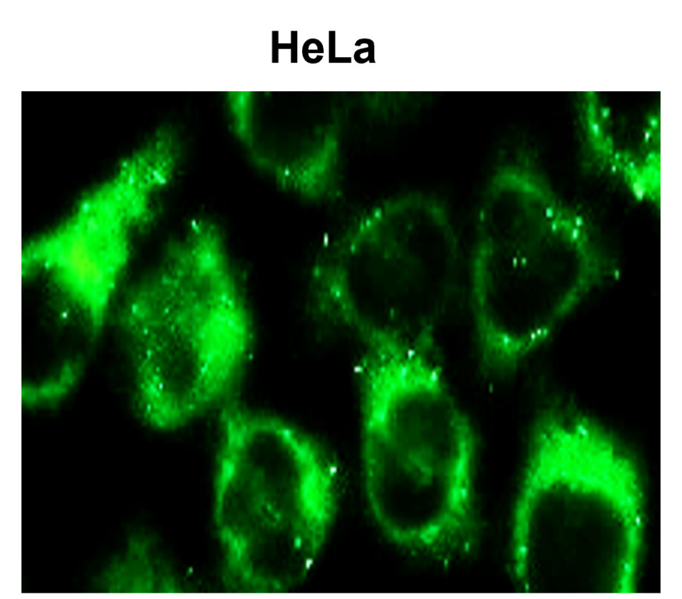
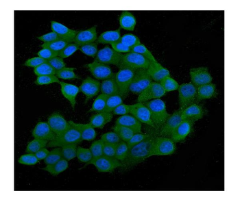
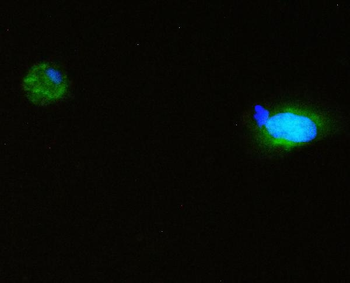
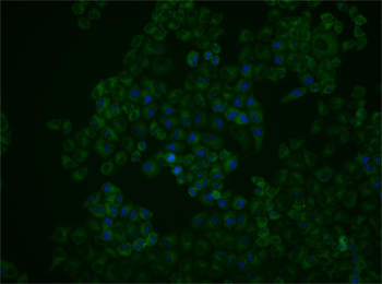
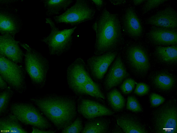
Immunofluorescence Validation of SQSTM1 In HeLa Cells. Immunofluorescent analysis of PFA-fixed HeLa cells labeling SQSTM1 with orb1240011 at 20 µg/mL, followed by goat anti-rabbit IgG secondary antibody at 1/1000 dilution (red) and Hoechst staining (blue). Alpha tubulin was stained with anti-alpha tubulin antibody following by goat anti-mouse IgG secondary antibody (green). Images were captured with confocal microscopy.
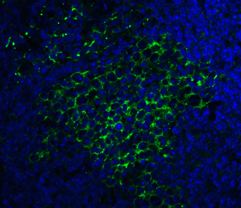
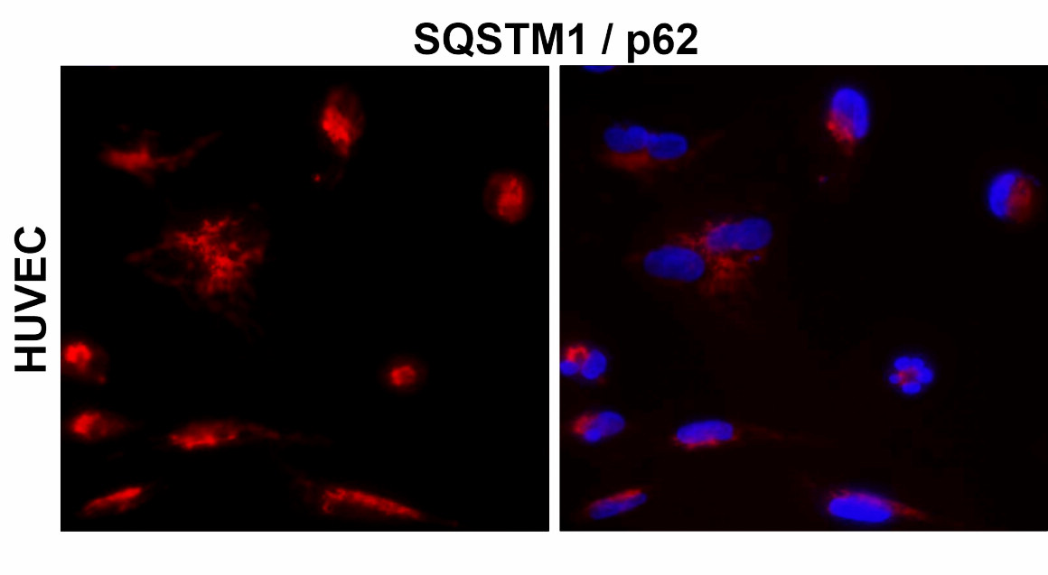
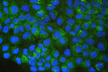
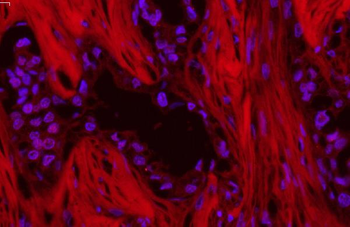
Immunofluorescence Validation of SQSTM1 in Mouse Spleen Tissue. Immunofluorescent analysis of 4% paraformaldehyde-fixed Mouse Spleen Tissue labeling SQSTM1 with orb1240011 at 20 µg/mL, followed by goat anti-rabbit IgG secondary antibody at 1/500 dilution (green) and DAPI staining (blue).
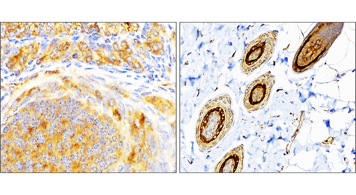
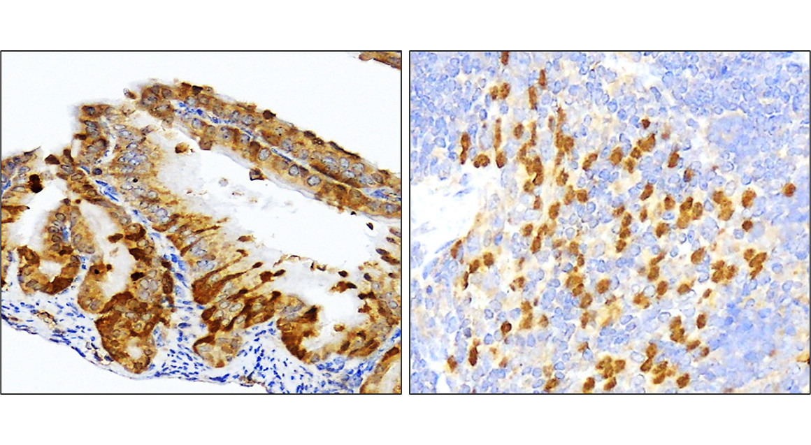
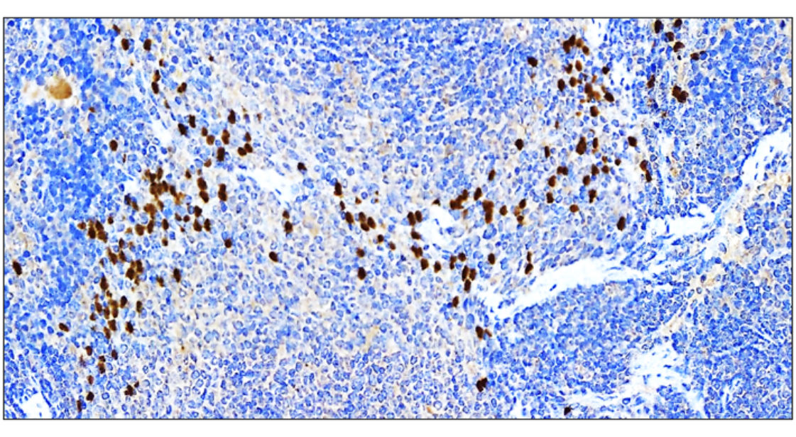
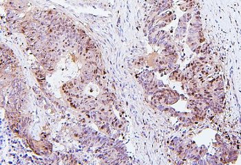
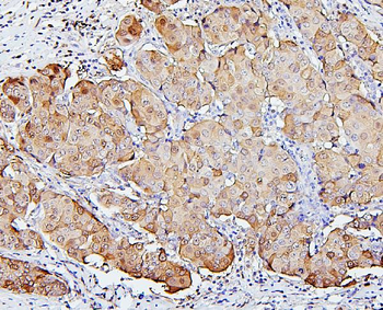
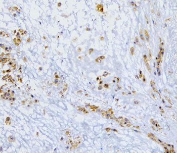
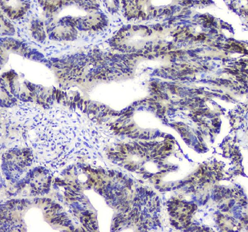
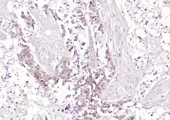
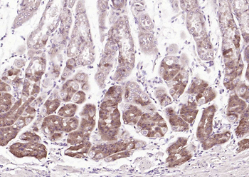
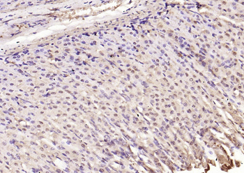
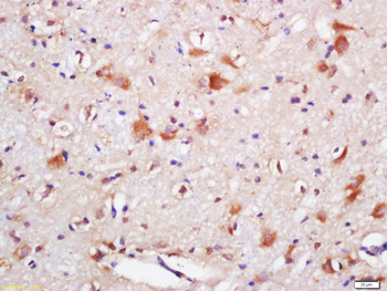
Immunohistochemistry Validation of SQSTM1 in Human Spleen Tissue. Immunohistochemical analysis of paraffin-emb edded Human Spleen Tissue using anti-SQSTM1 antibody (orb1240011) at 2 µg/mL. Tissue was fixed with formaldehyde and blocked with 10% serum for 1 h at RT; antigen retrieval was by heat mediation with a citrate buffer (pH6). Samples were incubated with primary antibody overnight at 4°C. A goat anti-rabbit IgG H&L (HRP) at 1/250 was used as secondary. Counter stained with Hematoxylin.
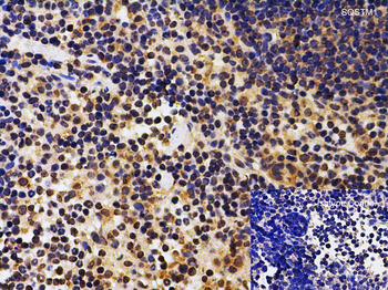
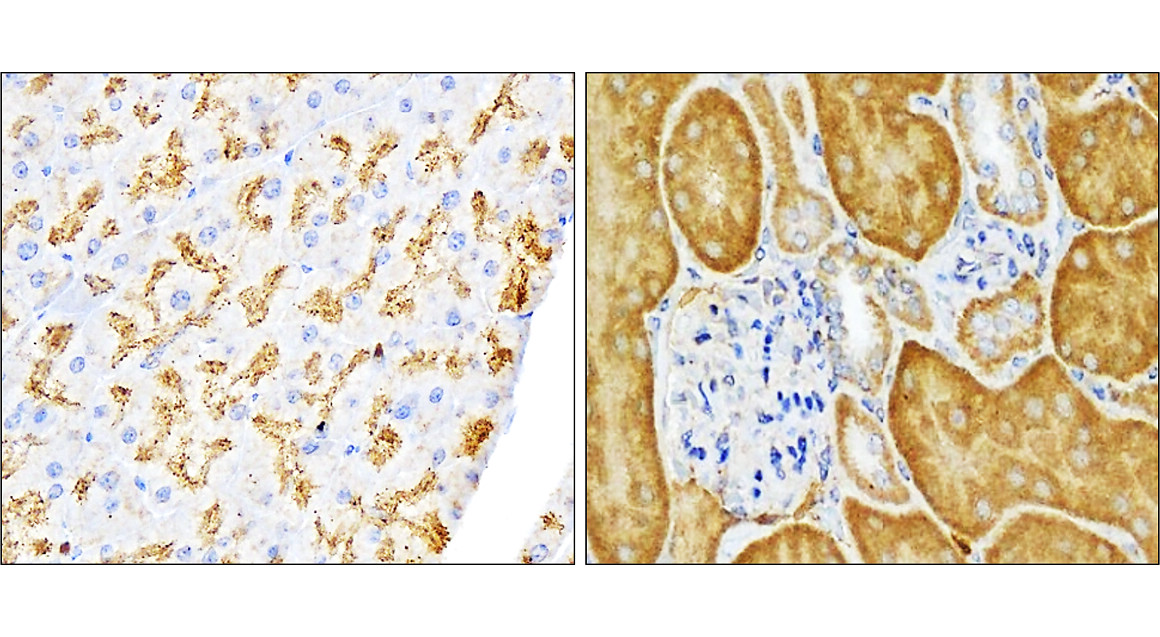
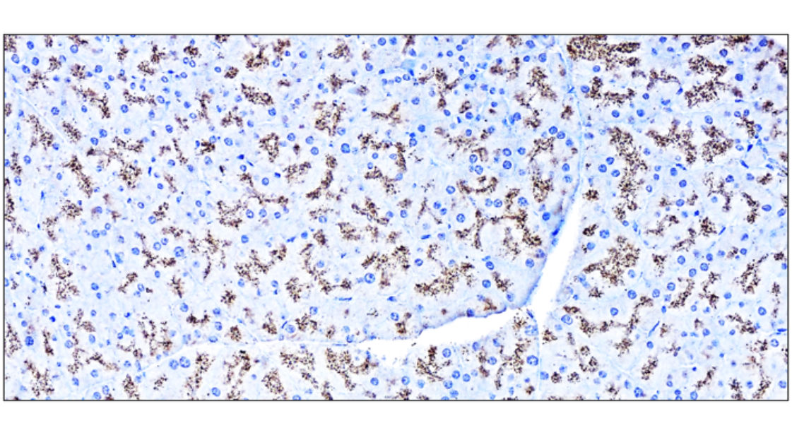
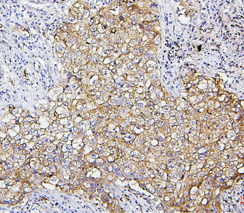
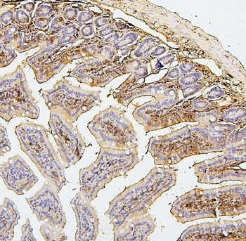
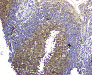
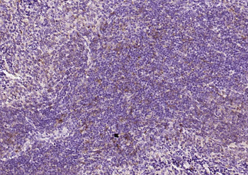
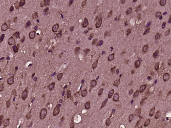
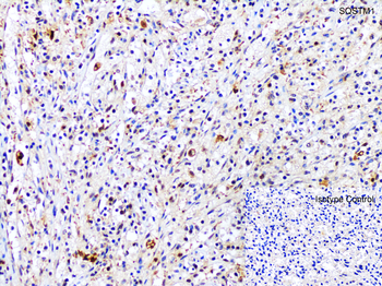
Immunohistochemistry Validation of SQSTM1 in Mouse Spleen Tissue. Immunohistochemical analysis of paraffin-embedded Mouse Spleen Tissue using anti-SQSTM1 antibody (orb1240011) at 2 µg/mL. Tissue was fixed with formaldehyde and blocked with 10% serum for 1 h at RT; antigen retrieval was by heat mediation with a citrate buffer (pH6). Samples were incubated with primary antibody overnight at 4°C. A goat anti-rabbit IgG H&L (HRP) at 1/250 was used as secondary. Counter stained with Hematoxylin.
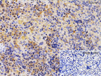
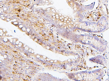
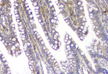
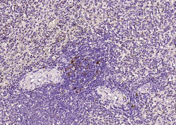
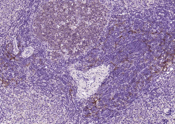
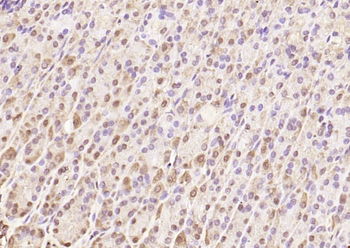
Immunohistochemistry Validation of SQSTM1 in Rat Spleen Tissue. Immunohistochemical analysis of paraffin-embedded Rat Spleen Tissue using anti-SQSTM1 antibody (orb1240011) at 2 µg/mL. Tissue was fixed with formaldehyde and blocked with 10% serum for 1 h at RT; antigen retrieval was by heat mediation with a citrate buffer (pH6). Samples were incubated with primary antibody overnight at 4°C. A goat anti-rabbit IgG H&L (HRP) at 1/250 was used as secondary. Counter stained with Hematoxylin.
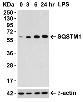
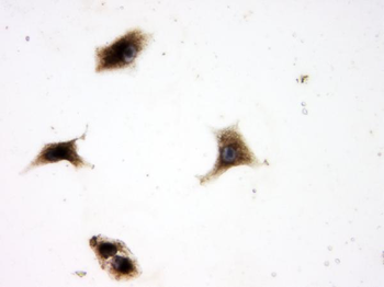
Induced Expression Validation in Mouse Macrophage Cells. Loading: 15 µg of lysates per lane. Antibodies: SQSTM1 orb1240011 (0.5 µg/mL), 1h incubation at RT in 5% NFDM/TBST. Secondary: Goat anti-rabbit IgG HRP conjugate at 1:10000 dilution. Raw 264.7 cells were treated with LPS (0.3 µg /mL) for different time period (0-24 hrs). There was an increase in SQSTM1 protein expression overtime after LPS treatment.
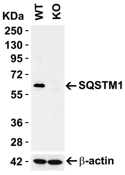
KO Validation in HEK293T Cells. Loading: 10 µg of HEK293T WT cell lysates or SQSTM1 KO cell lysates. Antibodies: SQSTM1 orb1240011 (1 µg/mL) and beta-actin orb1240312 (1 µg/mL), 1 h incubation at RT in 5% NFDM/TBST. Secondary: Goat Anti-Rabbit IgG HRP conjugate at 1:10000 dilution.
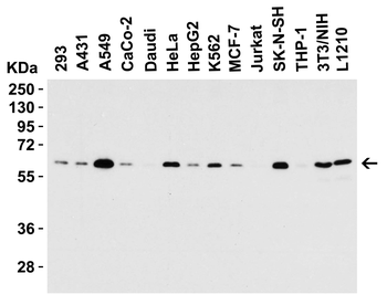
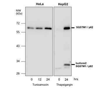
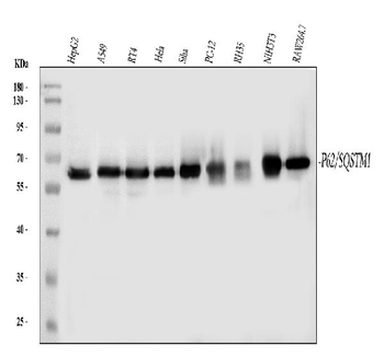
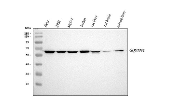
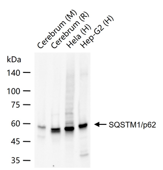
Western Blot Validation in Cell Lines. Loading: 15 µg of lysates per lane. Antibodies: SQSTM1 orb1240011 (0.5 µg/mL), 1 h incubation at RT in 5% NFDM/TBST. Secondary: Goat anti-rabbit IgG HRP conjugate at 1:10000 dilution.
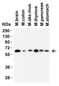
Western Blot Validation in Mouse Tissues. Loading: 15 µg of lysates per lane. Antibodies: SQSTM1 orb1240011 (1 µg/mL), 1h incubation at RT in 5% NFDM/TBST. Secondary: Goat anti-rabbit IgG HRP conjugate at 1:10000 dilution.
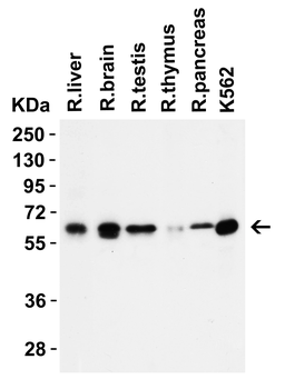
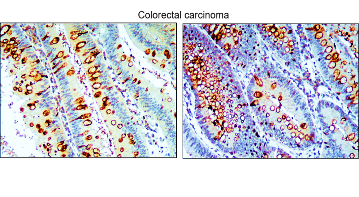
Western Blot Validation in Rat Tissues. Loading: 15 µg of lysates per lane. Antibodies: SQSTM1 orb1240011 (1 µg/mL), 1h incubation at RT in 5% NFDM/TBST. Secondary: Goat anti-rabbit IgG HRP conjugate at 1:10000 dilution.
Documents Download
Datasheet
Product Information
Request a Document
Protocol Information
WB
Western Blot (IB, immunoblot)
IHC-P
Immunohistochemistry Paraffin
IF
Immunofluorescence
ELISA
Enzyme-linked Immunosorbent Assay (EIA)
SQSTM1 Antibody (orb1240011)

- 0.0
Based on 0 reviews
Participating in our Biorbyt product reviews program enables you to support fellow scientists by sharing your firsthand experience with our products.
Login to Submit a ReviewAvailable Sizes
Select a size below
Free Secondary Antibody (20 ul)0/0
Please add an antibody product to your cart first.