You have no items in your shopping cart.
Description
Research Area
Metabolism Research
Images & Validation
−Item 1 of 2
| Tested Applications | ELISA, ICC, IF, WB |
|---|---|
| Dilution Range | WB (1:1000), ICC/IF (1:100) |
| Reactivity | Human, Mouse, Rat |
| Application Notes |
Key Properties
−| Host | Rabbit |
|---|---|
| Clonality | Polyclonal |
| Immunogen | Within 50 AA of N-terminal region of SOD1, unfolded beta barrel region |
| Target | SOD1 (UbetaB) |
| Purification | Peptide Affinity purified |
| Conjugation | APC |
Storage & Handling
−| Storage | Conjugated antibodies should be stored according to the product label |
|---|---|
| Buffer/Preservatives | 95.46mM Phosphate, 2.48mM MES and 2mM EDTA |
| Concentration | 1 mg/ml |
| Expiration Date | 12 months from date of receipt. |
| Disclaimer | For research use only |
Alternative Names
−SOD1 (UbetaB), SOD1, Superoxide dismutase [Cu-Zn], Cu/Zn superoxide dismutase, SODC_HUMAN, ALS1, USOD, SOD1 unfolded beta barrel region, Misfolded SOD1, Conformationally altered SOD1, Apo-SOD1, Superoxide dismutase 1, SOD
Quality Guarantee
Explore bioreagents carefree to elevate your research. All our products are rigorously tested for performance. If a product does not perform as described on its datasheet, our scientific support team will provide expert troubleshooting, a prompt replacement, or a refund. For full details, please see our Terms & Conditions and Buying Guide. Contact us at [email protected].
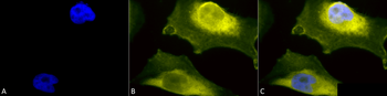
Immunocytochemistry/Immunofluorescence analysis using Rabbit Anti-SOD1 (UBB) Polyclonal Antibody. Tissue: Cervical cancer cell line (HeLa). Species: Human. Fixation: 2% Formaldehyde for 20 min at RT. Primary Antibody: Rabbit Anti-SOD1 (UBB) Polyclonal Antibody at 1:110 for 12 hours at 4°C. Secondary Antibody: R-PE Goat Anti-Rabbit (yellow) at 1:200 for 2 hours at RT. Counterstain: DAPI (blue) nuclear stain at 1:40000 for 2 hours at RT. Localization: Cytoplasm. Nucleus. Mitochondrion. Magnification: 100x. (A) DAPI (blue) nuclear stain. (B) Anti-SOD1 (UBB) Antibody. (C) Composite.
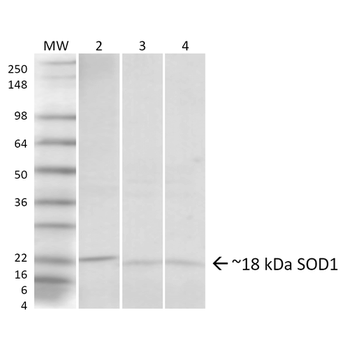
Western blot analysis of Human, Rat, Mouse Hela cells, Brain, Lung showing detection of ~18 kDa SOD1 (UBB) protein using Rabbit Anti-SOD1 (UBB) Polyclonal Antibody. Lane 1: Molecular Weight Ladder. Lane 2: Human HeLa Cell Lysates. Lane 3: Rat Brain. Lane 4: Mouse Lung. Load: 20 μg. Primary Antibody: Rabbit Anti-SOD1 (UBB) Polyclonal Antibody at 1:1000. Predicted/Observed Size: ~18 kDa.
UniProt Details
− No UniProt data available
NCBI Gene Details
− No NCBI Gene data available
NCBI Reference Sequences
−Associated Accession Numbers
Curated reference sequences for the gene transcript and protein product| RefSeq | CAG46542 |
|---|
Documents Download
Datasheet
Product Information
Request a Document
Protocol Information
WB
Western Blot (IB, immunoblot)
IF
Immunofluorescence
ICC
Immunocytochemistry
ELISA
Enzyme-linked Immunosorbent Assay (EIA)
SOD1 (UbetaB) Antibody (APC) (orb152253)

- 0.0
Based on 0 reviews
Participating in our Biorbyt product reviews program enables you to support fellow scientists by sharing your firsthand experience with our products.
Login to Submit a ReviewAvailable Sizes
Select a size below
Choose Conjugation or Carrier Free Version
Free Secondary Antibody (20 ul)0/0
Please add an antibody product to your cart first.