You have no items in your shopping cart.
Featured
Description
Images & Validation
−Item 1 of 7
| Tested Applications | ELISA, IF, IHC-P, WB |
|---|---|
| Reactivity | Human, Mouse, Rat |
| Predicted Reactivity | Bovine |
Key Properties
−| Antibody Type | Primary Antibody |
|---|---|
| Host | Rabbit |
| Clonality | Polyclonal |
| Isotype | IgG |
| Immunogen | Anti-SCF antibody (orb1240002) was raised against a peptide corresponding to 18 amino acids near the center of human SCF. The immunogen is located within amino acids 100-150 of SCF. |
| Target | KITLG |
| Molecular Weight | Predicted: 31kDObserved: 36 kD (31kD + 4 N-linked Glycosylations) |
| Purification | SCF Antibody is affinity chromatography purified via peptide column. |
| Conjugation | Unconjugated |
Storage & Handling
−| Storage | Maintain refrigerated at 2-8°C for up to 2 weeks. For long term storage store at -20°C in small aliquots to prevent freeze-thaw cycles. |
|---|---|
| Form/Appearance | Liquid |
| Buffer/Preservatives | SCF Antibody is supplied in PBS containing 0.02% sodium azide. |
| Concentration | 1 mg/mL |
| Expiration Date | 12 months from date of receipt. |
| Disclaimer | For research use only |
Alternative Names
−SCF Antibody: SF, MGF, SCF, FPH2, KL-1, Kitl, SHEP7, Kit ligand, Mast cell growth factor
Similar Products
−- Item 1 of 4
SCF Rabbit Polyclonal Antibody [orb11352]
FC, IF, IHC-Fr, IHC-P, WB
Goat
Human, Mouse, Rat
Rabbit
Polyclonal
Unconjugated
50 μl, 100 μl, 200 μl - Item 1 of 5
SCF/KITLG Rabbit Polyclonal Antibody [orb402451]
ICC, IHC, WB
Human, Mouse, Rat
Rabbit
Polyclonal
Unconjugated
100 μg - Item 1 of 1
- Item 1 of 1
- Item 1 of 1

Quality Guarantee
Explore bioreagents carefree to elevate your research. All our products are rigorously tested for performance. If a product does not perform as described on its datasheet, our scientific support team will provide expert troubleshooting, a prompt replacement, or a refund. For full details, please see our Terms & Conditions and Buying Guide. Contact us at [email protected].
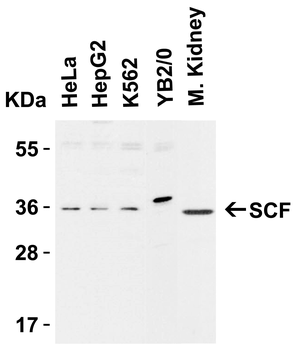
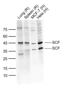
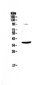
Western Blot Validation in Cell Lines and Tissues of Human, Mouse and Rat, Loading: 15 µg of lysates per lane. Antibodies: SCF orb1240002 (1 µg/mL), 1h incubation at RT in 5% NFDM/TBST. Secondary: Goat anti-rabbit IgG HRP conjugate at 1:10000 dilution.
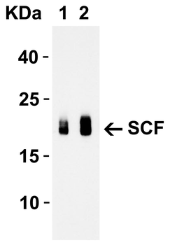
Western Blot Validation with Recombinant Protein, Loading: 30 ng of human SCF recombinant protein per lane. Antibodies: SCF orb1240002 (Lane 1: 1 µg/mL and Lane 2: 2 µg/mL), 1h incubation at RT in 5% NFDM/TBST. Secondary: Goat anti-rabbit IgG HRP conjugate at 1:10000 dilution. Observed at around 20kD.
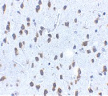
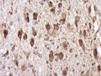
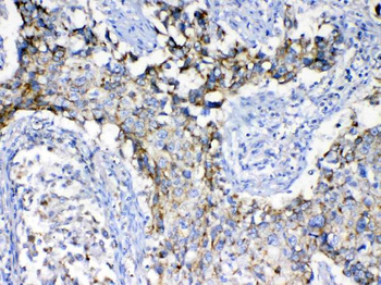
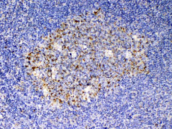
Immunohistochemistry Validation of SCF in Mouse Brain Tissue, Immunohistochemical analysis of paraffin-embedded mouse brain tissue using anti-SCF antibody (orb1240002) at 2.5 µg/mL. Tissue was fixed with formaldehyde and blocked with 10% serum for 1 h at RT; antigen retrieval was by heat mediation with a citrate buffer (pH6). Samples were incubated with primary antibody overnight at 4 °C. A goat anti-rabbit IgG H&L (HRP) at 1/250 was used as secondary. Counter stained with Hematoxylin.
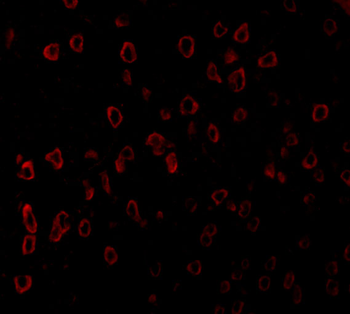
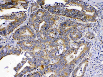
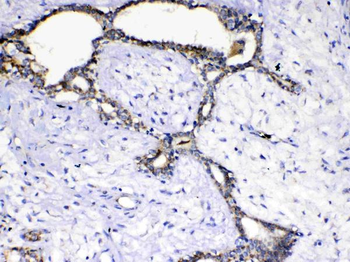
Immunofluorescence Validation of SCF in Human Brain Tissue, Immunofluorescent analysis of 4% paraformaldehyde-fixed human brain tissue labeling SCF with orb1240002 at 20 µg/mL, followed by goat anti-rabbit IgG secondary antibody at 1/500 dilution (red).
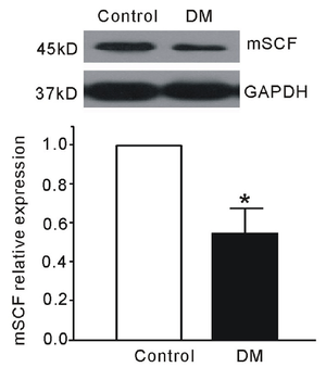
Regulation Validation of SCF in Streptozotocin (STZ)-induced Diabetic Mice, WB analysis showed protein expression level of SCF detected by anti-SCF antibody (orb1240002) in gastric smooth muscle was significantly decreased in STZ-induced diabetic mice as compared to the control.
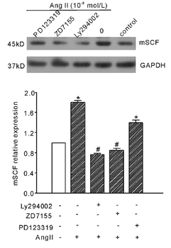
Regulated Expression Validation of SCF in Gastric Smooth Muscle Cells (GSMCs) of Mice, WB analysis of protein expression level of SCF detected by anti-SCF antibody (orb1240002). GSMCs were treated with PI3K inhibitor (LY294002), AT1R inhibitor (ZD7155) and AT2R inhibitor (PD123319) before Ang II treatment. Ang II (10^-8 mol/L) significantly increased SCF protein expression, which was reduced by treatment of LY294002 and ZD7155.
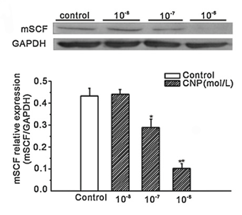
Regulated Expression Validation of SCF in GSMCs of Normal Mice, WB analysis of protein expression level of SCF detected by anti-SCF antibody (orb1240002). GSMCs were treated with C-type natriuretic peptide (CNP) at different doses for 48hr. CNP(10^-7 mol/L and 10^-6 mol/L) significantly decreased SCF protein expression level as compared to the control group.
Documents Download
Datasheet
Product Information
Request a Document
Protocol Information
WB
Western Blot (IB, immunoblot)
IHC-P
Immunohistochemistry Paraffin
IF
Immunofluorescence
ELISA
Enzyme-linked Immunosorbent Assay (EIA)
KITLG Antibody (orb1240002)

- 0.0
Based on 0 reviews
Participating in our Biorbyt product reviews program enables you to support fellow scientists by sharing your firsthand experience with our products.
Login to Submit a ReviewAvailable Sizes
Select a size below
Free Secondary Antibody (20 ul)0/0
Please add an antibody product to your cart first.