You have no items in your shopping cart.
Description
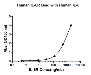
Images & Validation
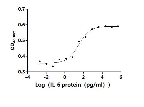
−| Application Notes |
|---|
Key Properties
−| Source | CHO Cell |
|---|---|
| Expression System | Eukaryotic cell |
| Biological Origin | Human |
| Endotoxins | ≤10 EU/mg |
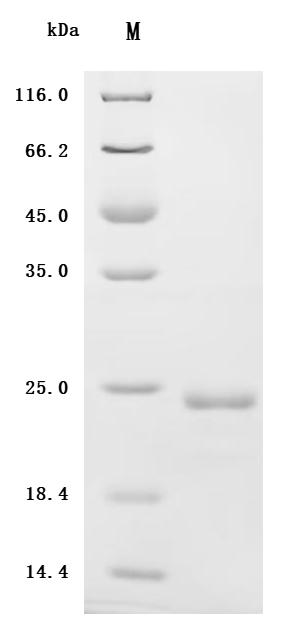
| MW | 20.64 kDa |
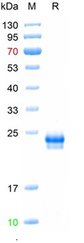
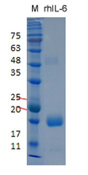
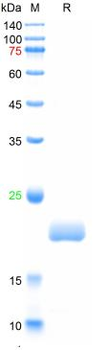
| Purity | ≥95% |
Storage & Handling
−| Storage | Use a manual defrost freezer and avoid repeated freeze - thaw cycles. 12 months from date of receipt, -20 to -70°C as supplied. 1 month, 2 to 8°C under sterile conditions after reconstitution. 6 months, -20 to -74°C under sterile conditions after reconstitution. |
|---|---|
| Form/Appearance | Lyophilized Powder |
| Expiration Date | 6 months from date of receipt. |
| Disclaimer | For research use only |
Alternative Names
−Interleukin 6, B-Cell Stimulatory Factor 2, B-Cell Differentiation Factor, CTL Differentiation Factor, Hybridoma Growth Factor, Interferon Beta-2, IFNB2, BSF-2, CDF, HGF, HSF
Similar Products
−- Item 1 of 2
Recombinant Human Interleukin-6 (IL6) Protein (Active) [orb1881858]
Greater than 95% as determined by SDS-PAGE
22.8 kDa
Yeast
1 mg, 100 μg, 20 μg 
Recombinant Human IL-6 Protein [orb2995250]
Unconjugated
SDS-PAGE: Greater than 95% as determined by reducing SDS-PAGE.
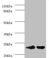
Predicted: 20.9 KDa. Observed: 20 KDa, reducing conditions
1 mg, 10 μg, 50 μg, 500 μg- Item 1 of 2
- Item 1 of 2
Recombinant human IL-6 protein, C-His (HEK293) [orb1516624]
> 90% as determined by SDS-PAGE method
20.8 kDa
20 μg, 100 μg, 500 μg - Item 1 of 2
Recombinant human IL-6 protein, C-His [orb1516828]
>95% as determined by SDS-PAGE
21 kDa
100 μg, 500 μg, 20 μg

Quality Guarantee
Explore bioreagents carefree to elevate your research. All our products are rigorously tested for performance. If a product does not perform as described on its datasheet, our scientific support team will provide expert troubleshooting, a prompt replacement, or a refund. For full details, please see our Terms & Conditions and Buying Guide. Contact us at [email protected].
Documents Download
Datasheet
Product Information
Request a Document
Protocol Information
Protein Handling and Storage Guide
Protein Handling Guide
Recombinant Human IL-6 (orb1921848)

- 0.0
Based on 0 reviews
Participating in our Biorbyt product reviews program enables you to support fellow scientists by sharing your firsthand experience with our products.
Login to Submit a ReviewAvailable Sizes
Select a size below