You have no items in your shopping cart.
Description
Research Area
Cell Biology
Images & Validation
−Item 1 of 7
| Tested Applications | FC, ICC, IHC, IP, WB |
|---|---|
| Dilution Range | Application : Flow-Cyt , ApplicationDilutionRange : Fixed in 4% formaldehyde and permeabilized with 90% methanol. 0.5 µl per 1 x 10^6 cells. , Application : ICC , ApplicationDilutionRange : 1:100 to 1:500. Epitope retrieval with citrate buffer pH 6.0 is recommended for FFPE cell sections. , Application : IHC , ApplicationDilutionRange : 1:100 to 1:500. Epitope retrieval with citrate buffer pH 6.0 is recommended for FFPE tissue sections. , Application : IP , ApplicationDilutionRange : 6 µl/mg lysate , Application : WB , ApplicationDilutionRange : 1:1000 |
| Reactivity | Human |
| Application Notes |
Key Properties
−| Antibody Type | Primary Antibody |
|---|---|
| Host | Rabbit |
| Clonality | Recombinant |
| Isotype | IgG |
| Clone No. | BLR283L |
| Immunogen | Between 1340 and C-terminus |
| Target | Met |
| Purification | Purified |
| Conjugation | Unconjugated |
Storage & Handling
−| Storage | Maintain refrigerated at 2-8°C for up to 2 weeks. For long term storage store at -20°C in small aliquots to prevent freeze-thaw cycles. |
|---|---|
| Form/Appearance | Liquid |
| Buffer/Preservatives | Borate Buffered Saline (BBS) pH 8.2 with 0.09% Sodium Azide, rAlbumin-Free |
| Concentration | 1000 µg/ml |
| Expiration Date | 12 months from date of receipt. |
| Disclaimer | For research use only |
Alternative Names
−AUTS9;c-Met;DFNB97;hepatocyte growth factor receptor;HGF receptor;HGF/SF receptor;HGFR;proto-oncogene c-Met;RCCP2;scatter factor receptor;SF receptor;tyrosine-protein kinase Met
Similar Products
−- Item 1 of 1
C-Met Recombinant Rabbit Monoclonal Antibody [orb1816867]
FC, WB
Mouse, Rat
Human
Rabbit
Recombinant
Unconjugated
50 μl, 100 μl, 25 μl - Item 1 of 1
Dnmt1 Recombinant Rabbit Monoclonal Antibody [orb2562712]
ICC, IF, IHC-Fr, IHC-P, WB
Human, Mouse, Rat
Human, Mouse, Rat
Rabbit
Recombinant
Unconjugated
50 μl, 100 μl, 25 μl 
Rabbit MACC1 Recombinant Monoclonal Antibody [orb1519659]
ICC, IHC, IP, WB
Human
Rabbit
Recombinant
Unconjugated
100 μg (BSA-free)Rabbit MACC1 Recombinant Monoclonal Antibody [orb1519660]
ICC, IHC, IP, WB
Human
Rabbit
Recombinant
Unconjugated
100 μl, 10 μlc-Met (Phospho-Y1349) Rabbit Monoclonal Antibody [orb2988907]
WB
Human
Rabbit
Monoclonal
Unconjugated
200 μl, 100 μl, 50 μl, 30 μl
Quality Guarantee
Explore bioreagents carefree to elevate your research. All our products are rigorously tested for performance. If a product does not perform as described on its datasheet, our scientific support team will provide expert troubleshooting, a prompt replacement, or a refund. For full details, please see our Terms & Conditions and Buying Guide. Contact us at [email protected].
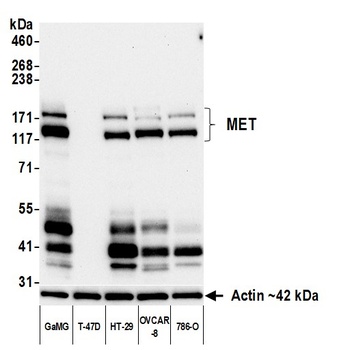
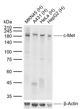
Detection of human MET by western blot. Samples: Whole cell lysate (10 µg) from GaMG, T-47D, HT-29, OVCAR-8, and 786-O cells prepared using NETN lysis buffer. Antibody: Rabbit anti-MET recombinant monoclonal antibody (orb1806454) used at 1:1000. Secondary: HRP-conjugated goat anti-rabbit IgG. Detection: Chemiluminescence with an exposure time of 30 seconds. Lower Panel: Rabbit anti-Actin recombinant monoclonal antibody.
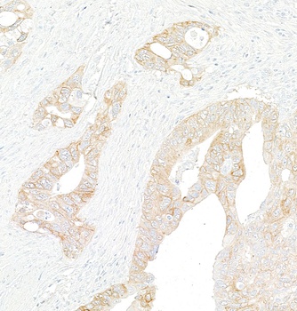
Detection of human MET by immunohistochemistry. Sample: FFPE section of colon carcinoma. Antibody: Rabbit anti-MET recombinant monoclonal antibody (orb1806454). Secondary: HRP-conjugated goat anti-rabbit IgG.
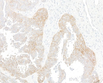
Detection of human MET by immunohistochemistry. Sample: FFPE section of ovarian carcinoma. Antibody: Rabbit anti-MET recombinant monoclonal antibody (orb1806454). Secondary: HRP-conjugated goat anti-rabbit IgG.
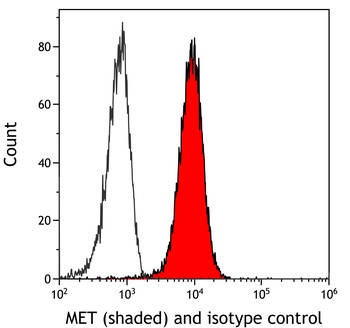
Detection of human MET (shaded) in HT-29 cells by flow cytometry. Antibody: Rabbit anti-MET recombinant monoclonal antibody (orb1806454) or isotype control (unshaded). Secondary: DyLight® 650-conjugated goat anti-rabbit IgG.
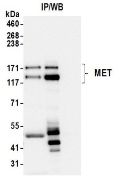
Detection of human MET by western blot of immunoprecipitates. Samples: Whole cell lysate (1 mg per IP; 10% of IP loaded) from GaMG cells prepared using NETN lysis buffer. Antibodies: Rabbit anti-MET recombinant monoclonal antibody (orb1806454) used for IP at 6 µl/mg lysate.

Detection of human MET by immunocytochemistry. Sample: FFPE section of GaMG cells. Antibody: Rabbit anti-MET recombinant monoclonal antibody (orb1806454). Secondary: HRP-conjugated goat anti-rabbit IgG.

Detection of human MET by immunocytochemistry. Sample: FFPE section of NCI-H226 cells. Antibody: Rabbit anti-MET recombinant monoclonal antibody (orb1806454). Secondary: HRP-conjugated goat anti-rabbit IgG.
Quick Database Links
UniProt Details
− No UniProt data available
NCBI Reference Sequences
−Associated Accession Numbers
Curated reference sequences for the gene transcript and protein product| Protein | NP_000236.2 |
|---|
Documents Download
Datasheet
Product Information
Request a Document
Protocol Information
WB
Western Blot (IB, immunoblot)
IHC
Immunohistochemistry
FC
Flow Cytometry
ICC
Immunocytochemistry
IP
Immunoprecipitation
Rabbit Met Recombinant Monoclonal Antibody (orb1806454)

- 0.0
Based on 0 reviews
Participating in our Biorbyt product reviews program enables you to support fellow scientists by sharing your firsthand experience with our products.
Login to Submit a ReviewAvailable Sizes
Select a size below
Choose Conjugation or Carrier Free Version
Free Secondary Antibody (20 ul)0/0
Please add an antibody product to your cart first.