You have no items in your shopping cart.
RAB3D Antibody (Biotin)
SKU: orb751914
Featured
Description
Images & Validation
−Item 1 of 2
| Tested Applications | WB |
|---|---|
| Dilution range | WB (1:1000) |
| Reactivity | Human, Mouse, Rat |
| Application Notes |
Key Properties
−| Host | Mouse |
|---|---|
| Clonality | Monoclonal |
| Isotype | IgG1 |
| Clone No. | 5000000000 |
| Immunogen | Full length recombinant RAB3D protein |
| Target | RAB3D |
| Molecular Weight | 24 kDa |
| Purification | Protein G Purified |
| Conjugation | Biotin |
Storage & Handling
−| Storage | Conjugated antibodies should be stored according to the product label |
|---|---|
| Buffer/Preservatives | 136.36mM Ethanolamine, 133.23 mM Chlorides, 9.55mM Phosphates, 9.55mM Sodium Bicarbonate |
| Concentration | 1mg/ml |
| Disclaimer | For research use only |
Alternative Names
−Member RAS Oncogene Family, D2-2, GOV, RAB16, RAD3D, Ras-Related Protein Rab-3D
Similar Products
−
Rab3A+Rab3B+Rab3C+Rab3D Rabbit Polyclonal Antibody (Biotin) [orb446696]
IF, IHC-Fr, IHC-P, WB
Human
Mouse, Rat
Rabbit
Polyclonal
Biotin
100 μlRAB3D Rabbit Polyclonal Antibody (Biotin) [orb2080984]
WB
Bovine, Human, Mouse, Rabbit, Rat
Rabbit
Polyclonal
Biotin
100 μl
Quality Guarantee
Explore bioreagents carefree to elevate your research. All our products are rigorously tested for performance. If a product does not perform as described on its datasheet, our scientific support team will provide expert troubleshooting, a prompt replacement, or a refund. For full details, please see our Terms & Conditions and Buying Guide. Contact us at [email protected].
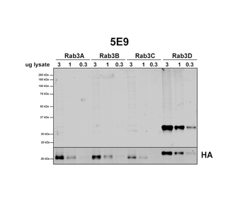
Western Blot analysis of Human HEK293 cells overexpressing RAB3A, RAB3B, RAB3C, and RAB3D showing detection of RAB3D protein using Mouse Anti-RAB3D Monoclonal Antibody, Clone 5E9. Lane 1: MW ladder. Lanes 2-4: HEK293 cells overexpressing RAB3A. Lanes 5-7: HEK293 cells overexpressing RAB3B. Lanes 8-10: HEK293 cells overexpressing RAB3C. Lanes 11-13: HEK293 cells overexpressing RAB3D. Primary Antibody: Mouse Anti-RAB3D Monoclonal Antibody.
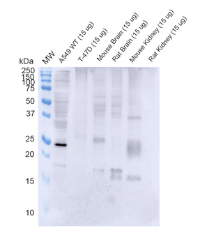
Western Blot analysis of Human, Mouse, Rat A549, T-47D cells, mouse and rat brain, kidney showing detection of RAB3D protein using Mouse Anti-RAB3D Monoclonal Antibody, Clone 5E9. Lane 1: MW ladder. Lane 2: WT A549 cells. Lane 3: T-47D cells. Lane 4: mouse brain. Lane 5: rat brain. Lane 6: mouse kidney. Lane 7: rat kidney. Load: 15 ug. Block: 5% Skim Milk Powder in TBST. Primary Antibody: Mouse Anti-RAB3D Monoclonal Antibody at 1:1000 for 2 hours at RT with shaking. Secondary Antibody: Goat anti-mouse IgG:HRP at 1:4000 for 1 hour at RT with shaking. Color Development: Chemiluminescent for HRP (Moss) for 5 min in RT.
UniProt Details
− No UniProt data available
NCBI Gene Details
− No NCBI Gene data available
NCBI Reference Sequences
−Associated Accession Numbers
Curated reference sequences for the gene transcript and protein product| Protein | NP_004274 |
|---|
Documents Download
Datasheet
Product Information
Request a Document
RAB3D Antibody (Biotin) (orb751914)

- 0.0
Based on 0 reviews
Participating in our Biorbyt product reviews program enables you to support fellow scientists by sharing your firsthand experience with our products.
Login to Submit a ReviewAvailable Sizes
Select a size below