You have no items in your shopping cart.
Featured
Description
Research Area
Signal Transduction
Images & Validation
−Item 1 of 4
| Tested Applications | AM, ICC, IF, IHC, WB |
|---|---|
| Dilution Range | WB (1:1000), IHC (1:1000), ICC/IF (1:100) |
| Reactivity | Bovine, Human, Mouse, Rat |
| Application Notes |
Key Properties
−| Host | Mouse |
|---|---|
| Clonality | Monoclonal |
| Isotype | IgG1 |
| Clone No. | 7000 |
| Immunogen | Recombinant rat PSD-95 |
| Target | PSD95 |
| Molecular Weight | 75kDa |
| Purification | Protein G Purified |
| Conjugation | APC |
Storage & Handling
−| Storage | Conjugated antibodies should be stored according to the product label |
|---|---|
| Buffer/Preservatives | 95.46mM Phosphate, 2.48mM MES and 2mM EDTA |
| Concentration | 1 mg/ml |
| Expiration Date | 12 months from date of receipt. |
| Disclaimer | For research use only |
Alternative Names
−PSD 95, PSD-95, PSD95, DLG4, SAP90, Synapse-associated protein 90, Postsynaptic density protein 95, Disks large homolog 4
Similar Products
−- Item 1 of 3
PSD95 Antibody (APC) [orb147006]
AM, ICC, IF, IHC, WB
Bovine, Human, Mouse, Rat
Mouse
Monoclonal
APC
100 μg 
DLGAP1 Rabbit Polyclonal Antibody (APC) [orb1005167]
ICC, IF
Bovine, Canine, Equine, Human, Mouse, Porcine, Rabbit, Rat, Sheep
Rabbit
Polyclonal
APC
100 μlGDA/GDC Rabbit Polyclonal Antibody (APC) [orb997442]
ICC, IF
Bovine, Canine, Equine, Human, Mouse, Rabbit, Rat
Rabbit
Polyclonal
APC
100 μlGDA/GDC Rabbit Polyclonal Antibody (APC-Cy7) [orb2501754]
ICC, IF
Bovine, Canine, Equine, Human, Mouse, Rabbit, Rat
Rabbit
Polyclonal
APC/Cy7
100 μlGDA/GDC Rabbit Polyclonal Antibody (APC-Cy5.5) [orb2501755]
ICC, IF
Bovine, Canine, Equine, Human, Mouse, Rabbit, Rat
Rabbit
Polyclonal
APC/Cy5.5
100 μl

Quality Guarantee
Explore bioreagents carefree to elevate your research. All our products are rigorously tested for performance. If a product does not perform as described on its datasheet, our scientific support team will provide expert troubleshooting, a prompt replacement, or a refund. For full details, please see our Terms & Conditions and Buying Guide. Contact us at [email protected].
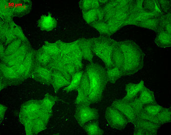
Immunohistochemistry analysis using Mouse Anti-PSD95 Monoclonal Antibody, Clone 7E3. Tissue: Neocortex. Species: Rat. Primary Antibody: Mouse Anti-PSD95 Monoclonal Antibody at 1:1000.
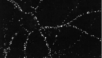
Immunocytochemistry/Immunofluorescence analysis using Mouse Anti-PSD95 Monoclonal Antibody, Clone 7E3. Tissue: HaCaT cells. Species: Human. Fixation: Cold 100% methanol for 10 minutes at -20°C. Primary Antibody: Mouse Anti-PSD95 Monoclonal Antibody at 1:100 for 1 hour at RT. Secondary Antibody: FITC Goat Anti-Mouse (green) at 1:50 for 1 hour at RT. Localization: Filamentous-like staining.
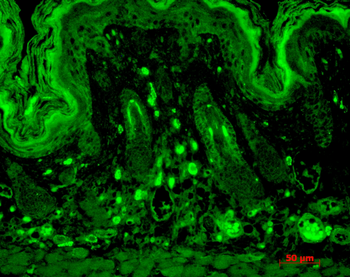
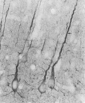
Immunohistochemistry analysis using Mouse Anti-PSD95 Monoclonal Antibody, Clone 7E3. Tissue: backskin. Species: Mouse. Fixation: Bouin's Fixative and paraffin-embedded. Primary Antibody: Mouse Anti-PSD95 Monoclonal Antibody at 1:100 for 1 hour at RT. Secondary Antibody: FITC Goat Anti-Mouse (green) at 1:50 for 1 hour at RT. Localization: Basal cell staining in the epidermis, some hair follicle staining, dermal staining. Backskin obtained from transgenic mice.
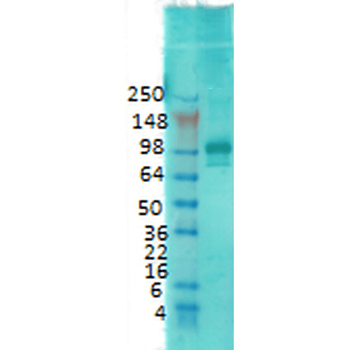
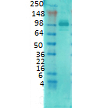
Western Blot analysis of Rat brain membrane lysate showing detection of PSD95 protein using Mouse Anti-PSD95 Monoclonal Antibody, Clone 7E3. Primary Antibody: Mouse Anti-PSD95 Monoclonal Antibody at 1:1000.
Quick Database Links
UniProt Details
− No UniProt data available
NCBI Gene Details
− No NCBI Gene data available
NCBI Reference Sequences
−Associated Accession Numbers
Curated reference sequences for the gene transcript and protein product| Protein | NP_062567.1 |
|---|
Documents Download
Datasheet
Product Information
Request a Document
Protocol Information
WB
Western Blot (IB, immunoblot)
IHC
Immunohistochemistry
IF
Immunofluorescence
ICC
Immunocytochemistry
PSD95 Antibody (APC) (orb147023)

- 0.0
Based on 0 reviews
Participating in our Biorbyt product reviews program enables you to support fellow scientists by sharing your firsthand experience with our products.
Login to Submit a ReviewAvailable Sizes
Select a size below
Choose Conjugation or Carrier Free Version
Free Secondary Antibody (20 ul)0/0
Please add an antibody product to your cart first.