You have no items in your shopping cart.
Featured
Description
Research Area
Cell Biology
Images & Validation
−Item 1 of 3
| Tested Applications | ELISA, IF, IHC-P, WB |
|---|---|
| Reactivity | Human, Mouse, Rat |
| Application Notes |
Key Properties
−| Antibody Type | Primary Antibody |
|---|---|
| Host | Rabbit |
| Clonality | Polyclonal |
| Isotype | IgG |
| Immunogen | PINK1 antibody was raised against a 16 amino acid peptide near the amino terminus of human PINK1.The immunogen is located within amino acids 120 - 170 of PINK1. |
| Target | PINK1 |
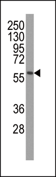
| Molecular Weight | Predicted: 55 kDa Observed: 53 kDa |
| Purification | PINK1 antibody is affinity chromatography purified via peptide column. |
| Conjugation | Unconjugated |
Storage & Handling
−| Storage | Maintain refrigerated at 2-8°C for up to 2 weeks. For long term storage store at -20°C in small aliquots to prevent freeze-thaw cycles. |
|---|---|
| Form/Appearance | Liquid |
| Buffer/Preservatives | PINK1 antibody is supplied in PBS containing 0.02% sodium azide. |
| Concentration | 1 mg/mL |
| Expiration Date | 12 months from date of receipt. |
| Disclaimer | For research use only |
Alternative Names
−PINK1 Antibody: BRPK, PARK6BRPK
Similar Products
−- Item 1 of 4
PINK1 Rabbit Polyclonal Antibody [orb331223]
IHC, WB
Bovine, Equine, Guinea pig, Mouse, Rabbit, Rat
Human
Rabbit
Polyclonal
Unconjugated
100 μl - Item 1 of 4
- Item 1 of 4
PINK1 Antibody (Ascites) [orb1440457]
IHC-P, WB
Human, Mouse
Mouse
Monoclonal
Unconjugated
50 μl, 100 μl - Item 1 of 1
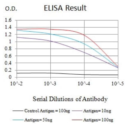
Human Serine/threonine-protein kinase PINK1, Mitochondrial (PINK1) ELISA Kit [orb1736595]
Human
0.32-20 ng/mL
0.14 ng/mL
48 T, 96 T - Item 1 of 5


Quality Guarantee
Explore bioreagents carefree to elevate your research. All our products are rigorously tested for performance. If a product does not perform as described on its datasheet, our scientific support team will provide expert troubleshooting, a prompt replacement, or a refund. For full details, please see our Terms & Conditions and Buying Guide. Contact us at [email protected].

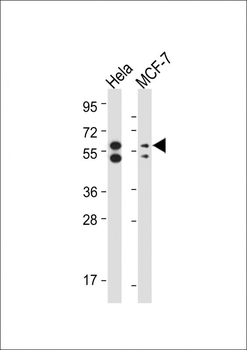
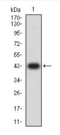
Western blot analysis of PINK1 in A431 cell lysate with PINK1 antibody at 1 µg/ml in (A) the absence and (B) the presence of blocking peptide.
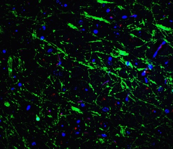
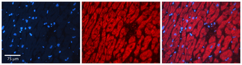
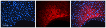
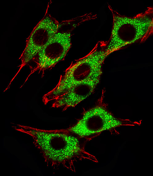
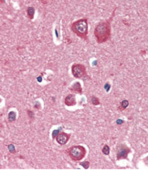
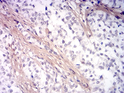
Immunofluorescence of PINK1 in mouse brain tissue with PINK1 Antibody at 20 µg/mL. Green: PINK1 antibody (orb1238781) Red: Phylloidin staining Blue: DAPI staining.
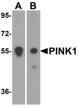
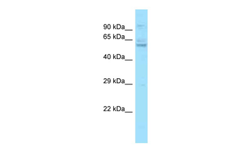
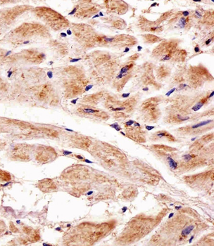
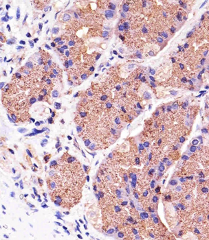
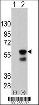
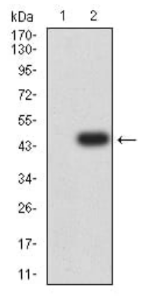
Western blot analysis of PINK1 in (A) mouse and (B) rat spleen tissue lysate with PINK1 antibody at 1 µg/mL.
Documents Download
Datasheet
Product Information
Request a Document
Protocol Information
WB
Western Blot (IB, immunoblot)
IHC-P
Immunohistochemistry Paraffin
IF
Immunofluorescence
ELISA
Enzyme-linked Immunosorbent Assay (EIA)
PINK1 Antibody (orb1238781)

- 0.0
Based on 0 reviews
Participating in our Biorbyt product reviews program enables you to support fellow scientists by sharing your firsthand experience with our products.
Login to Submit a ReviewAvailable Sizes
Select a size below
Free Secondary Antibody (20 ul)0/0
Please add an antibody product to your cart first.