You have no items in your shopping cart.
Phospho-EIF2S1 (Ser51) Recombinant Rabbit Monoclonal Antibody
SKU: orb1974725
Featured
Description
Research Area
Signal Transduction
Images & Validation
−Item 1 of 9
| Tested Applications | FC, ICC, IF, IHC-Fr, IHC-P, WB |
|---|---|
| Dilution Range | WB=1:500-2000, IHC-P=1:100-500, IHC-F=1:100-500, ICC/IF=1:100-200, IF=1:100-500, Flow-Cyt=1:100-200 |
| Reactivity | Human, Mouse, Rat |
| Predicted Reactivity | Rat |
Key Properties
−| Antibody Type | Primary Antibody |
|---|---|
| Host | Rabbit |
| Clonality | Recombinant |
| Isotype | IgG |
| Clone No. | B0D7 |
| Immunogen | A synthesized peptide derived from human eIF2A around the phosphorylation site of S51 EL-pS-RR |
| Target | EIF2S1 |
| Molecular Weight | 37 kDa |
| Purification | Affinity purified by Protein A |
| Conjugation | Unconjugated |
Storage & Handling
−| Storage | Maintain refrigerated at 2-8°C for up to 2 weeks. For long term storage store at -20°C in small aliquots to prevent freeze-thaw cycles. |
|---|---|
| Form/Appearance | Liquid |
| Buffer/Preservatives | 0.01M TBS (pH7.4) with 1% rAlbumin, 0.02% Proclin300 and 50% Glycerol. |
| Concentration | 1mg/ml |
| Expiration Date | 12 months from date of receipt. |
| Disclaimer | For research use only |
Alternative Names
−IF2A_HUMAN; EIF2S1; Eukaryotic translation initiation factor 2 subunit alpha (eIF-2-alpha | eIF-2A | eIF-2alpha | eIF2-alpha); EIF2A; IF2A_MOUSE; IF2A_RAT; eukaryotic translation initiation factor 2 subunit alpha; eukaryotic translation initiation factor 2, subunit 1 (alpha, 35kD ); eukaryotic translation initiation factor 2, subunit 1 alpha, 35kDa; EIF2S1 (phospho-S51); p-EIF2S1; phospho-EIF2S1; EIF2S1 (phospho-Ser51)
Quality Guarantee
Explore bioreagents carefree to elevate your research. All our products are rigorously tested for performance. If a product does not perform as described on its datasheet, our scientific support team will provide expert troubleshooting, a prompt replacement, or a refund. For full details, please see our Terms & Conditions and Buying Guide. Contact us at [email protected].
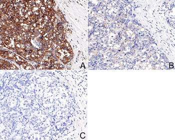
Immunohistochemical analysis of paraffin-embedded human breast carcinoma tissue with Rabbit anti-Phospho-EIF2S1 (S51) antibody (orb1974725) at 1/200 dilution. A: Untreated human breast carcinoma tissue, B: λ-PPase treated human breast carcinoma tissue, C: Negative control, The section was pre-treated using heat mediated antigen retrieval with Tris-EDTA buffer (pH9.0) for 20 minutes. The tissues were blocked in 1% BSA for 20 minutes at room temperature, washed with ddH2O and PBS, and then probed with the primary antibody (orb1974725) at 1/200 dilution for 1 hour at room temperature. The detection was performed using an HRP conjugated compact polymer system. DAB was used as the chromogen. Tissues were counterstained with hematoxylin and mounted with DPX.
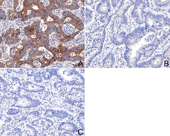
Immunohistochemical analysis of paraffin-embedded human colon carcinoma tissue with Rabbit anti-Phospho-EIF2S1 (S51) antibody (orb1974725) at 1/200 dilution. A: Untreated human colon carcinoma tissue, B: λ-PPase treated human colon carcinoma tissue, C: Negative control, The section was pre-treated using heat mediated antigen retrieval with Tris-EDTA buffer (pH9.0) for 20 minutes. The tissues were blocked in 1% BSA for 20 minutes at room temperature, washed with ddH2O and PBS, and then probed with the primary antibody (orb1974725) at 1/200 dilution for 1 hour at room temperature. The detection was performed using an HRP conjugated compact polymer system. DAB was used as the chromogen. Tissues were counterstained with hematoxylin and mounted with DPX.
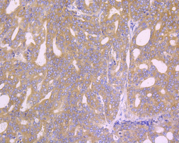
Immunohistochemical analysis of paraffin-embedded human liver tissue using anti-Phospho-EIF2S1 (S51) antibody. The section was pre-treated using heat mediated antigen retrieval with Tris-EDTA buffer (pH 8.0-8.4) for 20 minutes. The tissues were blocked in 5% BSA for 30 minutes at room temperature, washed with ddH2O and PBS, and then probed with the primary antibody (orb1974725, 1/50) for 30 minutes at room temperature. The detection was performed using an HRP conjugated compact polymer system. DAB was used as the chromogen. Tissues were counterstained with hematoxylin and mounted with DPX.
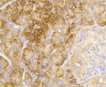
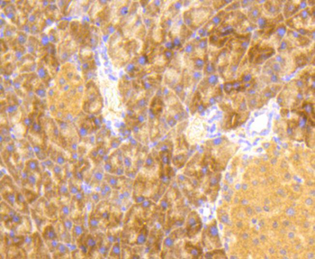
Immunohistochemical analysis of paraffin-embedded human pancreas tissue using anti-Phospho-EIF2S1 (S51) antibody. The section was pre-treated using heat mediated antigen retrieval with Tris-EDTA buffer (pH 8.0-8.4) for 20 minutes. The tissues were blocked in 5% BSA for 30 minutes at room temperature, washed with ddH2O and PBS, and then probed with the primary antibody (orb1974725, 1/50) for 30 minutes at room temperature. The detection was performed using an HRP conjugated compact polymer system. DAB was used as the chromogen. Tissues were counterstained with hematoxylin and mounted with DPX.
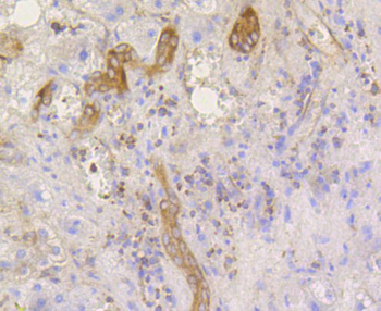
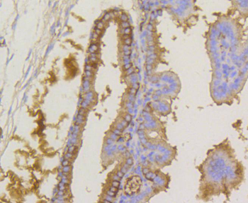
Immunohistochemical analysis of paraffin-embedded human prostate carcinoma tissue using anti-Phospho-EIF2S1 (S51) antibody. The section was pre-treated using heat mediated antigen retrieval with Tris-EDTA buffer (pH 8.0-8.4) for 20 minutes. The tissues were blocked in 5% BSA for 30 minutes at room temperature, washed with ddH2O and PBS, and then probed with the primary antibody (orb1974725, 1/50) for 30 minutes at room temperature. The detection was performed using an HRP conjugated compact polymer system. DAB was used as the chromogen. Tissues were counterstained with hematoxylin and mounted with DPX.
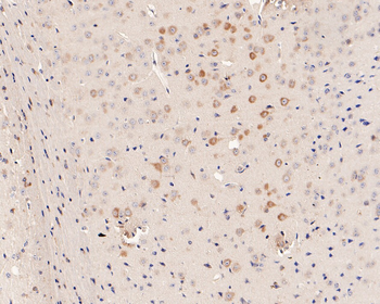
Immunohistochemical analysis of paraffin-embedded mouse brain tissue with Rabbit anti-Phospho-EIF2S1 (S51) antibody (orb1974725) at 1/200 dilution. The section was pre-treated using heat mediated antigen retrieval with Tris-EDTA buffer (pH9.0) for 20 minutes. The tissues were blocked in 1% BSA for 20 minutes at room temperature, washed with ddH2O and PBS, and then probed with the primary antibody (orb1974725) at 1/200 dilution for 1 hour at room temperature. The detection was performed using an HRP conjugated compact polymer system. DAB was used as the chromogen. Tissues were counterstained with hematoxylin and mounted with DPX.
Immunohistochemical analysis of paraffin-embedded mouse pancreas tissue using anti-Phospho-EIF2S1 (S51) antibody. The section was pre-treated using heat mediated antigen retrieval with Tris-EDTA buffer (pH 8.0-8.4) for 20 minutes. The tissues were blocked in 5% BSA for 30 minutes at room temperature, washed with ddH2O and PBS, and then probed with the primary antibody (orb1974725, 1/50) for 30 minutes at room temperature. The detection was performed using an HRP conjugated compact polymer system. DAB was used as the chromogen. Tissues were counterstained with hematoxylin and mounted with DPX.
Immunohistochemical analysis of paraffin-embedded mouse placenta tissue using anti-Phospho-EIF2S1 (S51) antibody. The section was pre-treated using heat mediated antigen retrieval with Tris-EDTA buffer (pH 8.0-8.4) for 20 minutes. The tissues were blocked in 5% BSA for 30 minutes at room temperature, washed with ddH2O and PBS, and then probed with the primary antibody (orb1974725, 1/50) for 30 minutes at room temperature. The detection was performed using an HRP conjugated compact polymer system. DAB was used as the chromogen. Tissues were counterstained with hematoxylin and mounted with DPX.
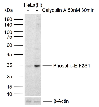
Sample: Lane 1: Human HeLa cell lysates, Lane 2: Human HeLa cells treated with Calyculin A 50nM 30 min, Primary: Anti-Phospho-EIF2S1 (Ser51) (orb1974725) at 1/2000 dilution, Secondary: IRDye800CW Goat Anti-Rabbit IgG at 1/20000 dilution, Predicted band size: 36 kDa, Observed band size: 35 kDa.
Quick Database Links
Gene Symbol
EIF2S1
UniProt
UniProt Details
− No UniProt data available
Documents Download
Datasheet
Product Information
Request a Document
Protocol Information
WB
Western Blot (IB, immunoblot)
IHC-P
Immunohistochemistry Paraffin
IHC-Fr
Immunohistochemistry Frozen
FC
Flow Cytometry
IF
Immunofluorescence
ICC
Immunocytochemistry
Phospho-EIF2S1 (Ser51) Recombinant Rabbit Monoclonal Antibody (orb1974725)

- 0.0
Based on 0 reviews
Participating in our Biorbyt product reviews program enables you to support fellow scientists by sharing your firsthand experience with our products.
Login to Submit a ReviewAvailable Sizes
Select a size below
Free Secondary Antibody (20 ul)0/0
Please add an antibody product to your cart first.