You have no items in your shopping cart.
Phospho-Caseine Kinase 1 alpha (Tyr294) Rabbit Polyclonal Antibody
SKU: orb156273
Featured
Description
Research Area
Signal Transduction
Images & Validation
−Item 1 of 2
| Tested Applications | IF, IHC-Fr, IHC-P, WB |
|---|---|
| Dilution Range | WB=1:500-2000, IHC-P=1:100-500, IHC-F=1:100-500, IF=1:100-500 |
| Reactivity | Human, Rat |
| Predicted Reactivity | Gallus, Mouse, Rabbit, Sheep |
Related Conjugates & Formulations
−Key Properties
−| Antibody Type | Primary Antibody |
|---|---|
| Host | Rabbit |
| Clonality | Polyclonal |
| Isotype | IgG |
| Immunogen | KLH conjugated synthesised phosphopeptide derived from human Caseine Kinase 1 alpha around the phosphorylation site of Tyr294 YD(p-Y)TF |
| Target | CSNK1A1 |
| Molecular Weight | 39 kDa |
| Purification | Affinity purified by Protein A |
| Conjugation | Unconjugated |
Storage & Handling
−| Storage | Maintain refrigerated at 2-8°C for up to 2 weeks. For long term storage store at -20°C in small aliquots to prevent freeze-thaw cycles. |
|---|---|
| Form/Appearance | Liquid |
| Buffer/Preservatives | 0.01M TBS (pH7.4) with 1% rAlbumin, 0.02% Proclin300 and 50% Glycerol. |
| Concentration | 1mg/ml |
| Expiration Date | 12 months from date of receipt. |
| Disclaimer | For research use only |
Alternative Names
−CK1; CK1a; CKIa; HEL-S-77p; HLCDGP1; PRO2975; KC1A_HUMAN; CSNK1A1; CKI-alpha; 2.7.11.1; casein kinase 1 alpha 1; casein kinase 1, alpha 1; clock regulator kinase; CSNK1A1 | CKI-alpha (phospho-Y294); p-CKI-alpha; phospho-CKI-alpha; p-CK1; CSNK1A1 | CKI-alpha (phospho-Tyr294)
Similar Products
−
Phospho-Caseine Kinase 1 alpha (Tyr294) Rabbit Polyclonal Antibody (PE-Cy7) [orb922091]
IF
Gallus, Mouse, Rabbit, Sheep
Human, Rat
Rabbit
Polyclonal
PE/Cy7
100 μlPhospho-Caseine Kinase 1 alpha (Tyr294) Rabbit Polyclonal Antibody (PE-Cy5.5) [orb922061]
IF
Gallus, Mouse, Rabbit, Sheep
Human, Rat
Rabbit
Polyclonal
PE/Cy5.5
100 μlPhospho-Caseine Kinase 1 alpha (Tyr294) Rabbit Polyclonal Antibody (Cy5) [orb920261]
IF
Gallus, Mouse, Rabbit, Sheep
Human, Rat
Rabbit
Polyclonal
Cy5
100 μlPhospho-Caseine Kinase 1 alpha (Tyr294) Rabbit Polyclonal Antibody (AP) [orb920232]
IHC-Fr, IHC-P, WB
Gallus, Mouse, Rabbit, Sheep
Human, Rat
Rabbit
Polyclonal
AP
100 μlPhospho-Caseine Kinase 1 alpha (Tyr294) Rabbit Polyclonal Antibody (PE) [orb505523]
IF
Gallus, Mouse, Rabbit, Sheep
Human, Rat
Rabbit
Polyclonal
PE
100 μl
Quality Guarantee
Explore bioreagents carefree to elevate your research. All our products are rigorously tested for performance. If a product does not perform as described on its datasheet, our scientific support team will provide expert troubleshooting, a prompt replacement, or a refund. For full details, please see our Terms & Conditions and Buying Guide. Contact us at [email protected].
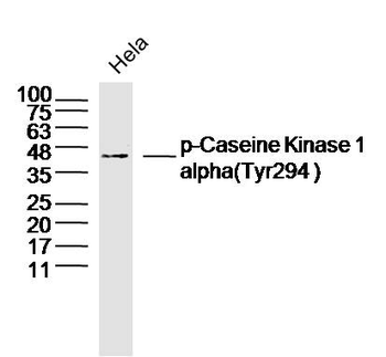
Sample: Hela Cell (Human) Lysate at 40 ug, Primary: Anti-phospho-Caseine Kinase 1 alpha (Tyr294) (orb156273) at 1/300 dilution, Secondary: IRDye800CW Goat Anti-Rabbit IgG at 1/20000 dilution, Predicted band size: 39 kD, Observed band size: 39 kD.
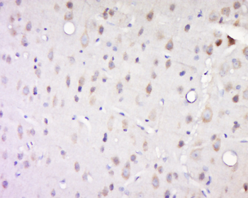
Tissue/Cell: rat brain tissue, 4% Paraformaldehyde-fixed and paraffin-embedded, Antigen retrieval: citrate buffer (0.01M, pH 6.0), Boiling bathing for 15 min, Block endogenous peroxidase by 3% Hydrogen peroxide for 30 min, Blocking buffer (normal goat serum) at 37°C for 20 min, Incubation: Anti-phospho-Caseine Kinase 1 alpha (Tyr294) Polyclonal Antibody, Unconjugated (orb156273) 1:500, overnight at 4°C, followed by conjugation to the secondary antibody and DAB staining.
Quick Database Links
Gene Symbol
CSNK1A1
UniProt
UniProt Details
− No UniProt data available
Documents Download
Datasheet
Product Information
Request a Document
Protocol Information
WB
Western Blot (IB, immunoblot)
IHC-P
Immunohistochemistry Paraffin
IHC-Fr
Immunohistochemistry Frozen
IF
Immunofluorescence
Phospho-Caseine Kinase 1 alpha (Tyr294) Rabbit Polyclonal Antibody (orb156273)

- 0.0
Based on 0 reviews
Participating in our Biorbyt product reviews program enables you to support fellow scientists by sharing your firsthand experience with our products.
Login to Submit a ReviewAvailable Sizes
Select a size below
Free Secondary Antibody (20 ul)0/0
Please add an antibody product to your cart first.