You have no items in your shopping cart.
Featured
Description
Research Area
Coactivators and Corepressors, DNA and RNA Research, Epigenetics and Nuclear Signaling, Hormone Research, Signal Transduction
Images & Validation
−Item 1 of 5
| Tested Applications | FC, ICC, WB |
|---|---|
| Dilution Range | WB=1:500-2000, ICC/IF=1:100-500, Flow-Cyt=2ug/Test |
| Reactivity | Human, Mouse, Rat |
| Predicted Reactivity | Bovine, Equine, Sheep |
Related Conjugates & Formulations
−Key Properties
−| Antibody Type | Primary Antibody |
|---|---|
| Host | Rabbit |
| Clonality | Polyclonal |
| Isotype | IgG |
| Immunogen | KLH conjugated synthetic peptide derived from human PHB2 (281-299/299aa) |
| Target | PHB2 |
| Molecular Weight | 35 kDa |
| Purification | Affinity purified by Protein A |
| Conjugation | Unconjugated |
Storage & Handling
−| Storage | Maintain refrigerated at 2-8°C for up to 2 weeks. For long term storage store at -20°C in small aliquots to prevent freeze-thaw cycles. |
|---|---|
| Form/Appearance | Liquid |
| Buffer/Preservatives | 0.01M TBS (pH7.4) with 1% rAlbumin, 0.02% Proclin300 and 50% Glycerol. |
| Concentration | 1mg/ml |
| Expiration Date | 12 months from date of receipt. |
| Disclaimer | For research use only |
Alternative Names
−BAP; BCAP37; Bap37; PNAS-141; REA; hBAP; p22; Bap-37; Bcap27; PHB2_HUMAN; PHB2; B-cell receptor-associated protein BAP37; D-prohibitin; Repressor of estrogen receptor activity; PHB2_MOUSE; PHB2_RAT; B-cell receptor-associated protein BAP37 (BAP-37);
Similar Products
−- Item 1 of 8
REA/PHB2 Rabbit Polyclonal Antibody [orb669110]
ELISA, FC, ICC, IF, IHC, WB
Human, Mouse, Rat
Rabbit
Polyclonal
Unconjugated
100 μg - Item 1 of 4
PHB2 Antibody (Y248) [orb1929408]
IHC-P, WB
Bovine
Human, Mouse, Rat
Rabbit
Polyclonal
Unconjugated
50 μl, 100 μl - Item 1 of 4
PHB2 Rabbit Polyclonal Antibody [orb630234]
ELISA, IF, IHC, IP, WB
Human, Mouse, Rat
Rabbit
Polyclonal
Unconjugated
50 μg, 100 μg - Item 1 of 3
PHB2 Antibody (Y128) [orb1929409]
IHC-P, WB
Rat
Human, Mouse
Rabbit
Polyclonal
Unconjugated
50 μl, 100 μl - Item 1 of 3
REA (PHB2) Antibody (C-term) [orb1929410]
IHC-P, WB
Mouse, Rat
Human
Rabbit
Polyclonal
Unconjugated
100 μl, 50 μl


Quality Guarantee
Explore bioreagents carefree to elevate your research. All our products are rigorously tested for performance. If a product does not perform as described on its datasheet, our scientific support team will provide expert troubleshooting, a prompt replacement, or a refund. For full details, please see our Terms & Conditions and Buying Guide. Contact us at [email protected].
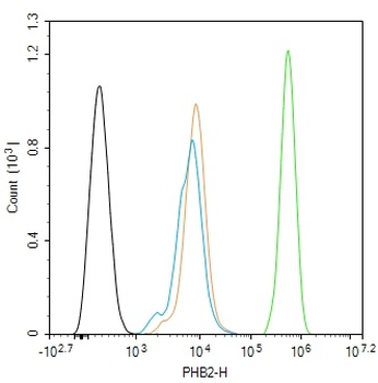
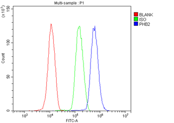
Blank control (black line): Hela. Primary Antibody (green line): Rabbit Anti-PHB2 antibody (orb185934), dilution: 2 ug/Test, Secondary Antibody (white blue line): Goat anti-rabbit IgG-FITC, dilution: 0.5 ug/Test. Isotype control (orange line): Normal Rabbit IgG, Protocol, The cells were fixed with 4% PFA (10 min at room temperature) and then permeabilized with 90% ice-cold methanol for 20 min at -20°C, The cells were then incubated in 5% BSA to block non-specific protein-protein interactions for 30 min at room temperature. Cells stained with Primary Antibody for 30 min at room temperature. The secondary antibody used for 40 min at room temperature. Acquisition of 20000 events was performed.
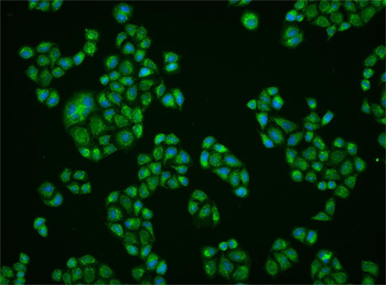
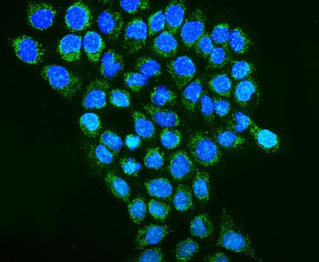
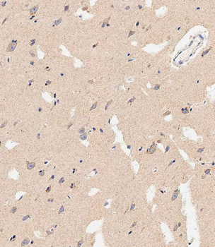
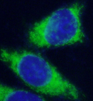
Hela cell, 4% Paraformaldehyde-fixed, Triton X-100 at room temperature for 20 min, Blocking buffer (normal goat serum) at 37°C for 20 min, Antibody incubation with (PHB2) polyclonal Antibody, Unconjugated (orb185934) 1:50, 90 minutes at 37°C, followed by a conjugated Goat Anti-Rabbit IgG antibody at 37°C for 90 minutes, DAPI (blue) was used to stain the cell nuclei.
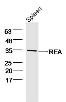
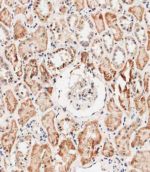
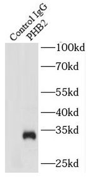
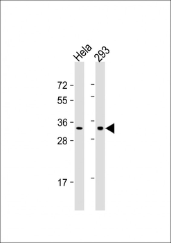
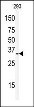
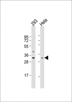
Protein: spleen (mouse) lysate at 40 ug, Primary: rabbit Anti-REA (orb185934) at 1:300, Secondary: HRP conjugated Goat-Anti-rabbit IgG (orb572747) at 1:5000, Predicted band size: 33 kD, Observed band size: 35 kD.
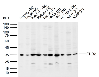
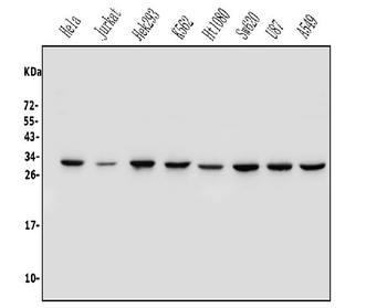
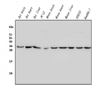
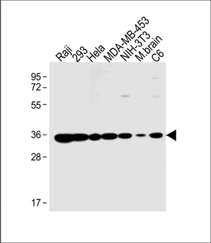
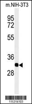
Sample: Lane 1: Mouse Kidney tissue lysates, Lane 2: Mouse Testis tissue lysates, Lane 3: Mouse NIH/3T3 cell lysates, Lane 4: Rat Kidney tissue lysates, Lane 5: Rat Testis tissue lysates, Lane 6: Human HeLa cell lysates, Lane 7: Human 293T cell lysates, Lane 8: Human HT-1080 cell lysates, Lane 9: Human A431 cell lysates, Lane 10: Human A549 cell lysates, Primary: Anti-PHB2 (orb185934) at 1/1000 dilution, Secondary: IRDye800CW Goat Anti-Rabbit IgG at 1/20000 dilution, Predicted band size: 33 kDa, Observed band size: 35 kDa.
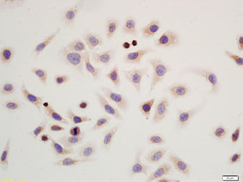
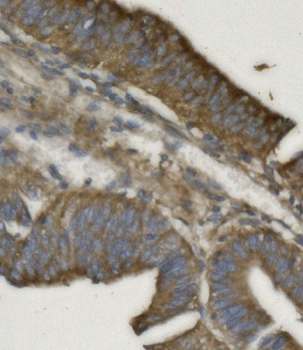
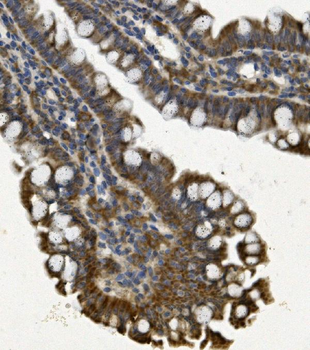
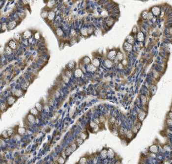
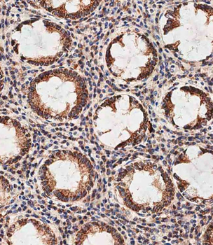
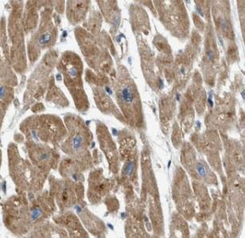
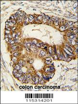
Tissue/Cell: colonic epithelial cells, Block endogenous peroxidase by 3% Hydrogen peroxide for 30 min, Blocking buffer (normal goat serum) at 37°C for 20 min, Incubation: Anti-REA Polyclonal Antibody, Unconjugated (orb185934) 1:100, overnight at 4°C, followed by conjugation to the secondary antibody and DAB staining.
Quick Database Links
Gene Symbol
PHB2
UniProt
UniProt Details
− No UniProt data available
Documents Download
Datasheet
Product Information
Request a Document
Protocol Information
WB
Western Blot (IB, immunoblot)
FC
Flow Cytometry
ICC
Immunocytochemistry
PHB2 Rabbit Polyclonal Antibody (orb185934)

- 0.0
Based on 0 reviews
Participating in our Biorbyt product reviews program enables you to support fellow scientists by sharing your firsthand experience with our products.
Login to Submit a ReviewAvailable Sizes
Select a size below
Free Secondary Antibody (20 ul)0/0
Please add an antibody product to your cart first.