You have no items in your shopping cart.
Featured
KO/KD
Validated
Validated
Description
Research Area
Immunology & Inflammation
Images & Validation
−Item 1 of 10
| Tested Applications | ELISA, IF, IHC-P, KO/KD Validated, WB |
|---|---|
| Reactivity | Human, Mouse |
| Predicted Reactivity | Rat |
Key Properties
−| Antibody Type | Primary Antibody |
|---|---|
| Host | Rabbit |
| Clonality | Polyclonal |
| Isotype | IgG |
| Immunogen | Anti-PD-L2 antibody (orb1239861) was raised against a peptide corresponding to 16 amino acids near the center of human PD-L2. The immunogen is located within amino acids 140-190 of PD-L2. |
| Target | PDCD1LG2 |
| Molecular Weight | Predicted: 31kDObserved: 29 kD |
| Purification | PD-L2 Antibody is affinity chromatography purified via peptide column. |
| Conjugation | Unconjugated |
Storage & Handling
−| Storage | Maintain refrigerated at 2-8°C for up to 2 weeks. For long term storage store at -20°C in small aliquots to prevent freeze-thaw cycles. |
|---|---|
| Form/Appearance | Liquid |
| Buffer/Preservatives | PD-L2 Antibody is supplied in PBS containing 0.02% sodium azide. |
| Concentration | 1 mg/mL |
| Expiration Date | 12 months from date of receipt. |
| Disclaimer | For research use only |
Alternative Names
−PD-L2 Antibody: B7DC, Btdc, PDL2, CD273, PD-L2, PDCD1L2, bA574F11.2, B7DC, Programmed cell death 1 ligand 2, Butyrophilin B7-DC, PD-1 ligand 2
Similar Products
−- Item 1 of 9
- Item 1 of 8
PDCD1LG2 Antibody [orb1239853]
ELISA, FC, ICC, IF, IHC-P, WB
Human
Mouse
Monoclonal
Unconjugated
0.1 mg, 0.02 mg - Item 1 of 8
PDCD1LG2 Antibody [orb1239860]
ELISA, FC, ICC, IF, IHC-P, WB
Human
Mouse
Monoclonal
Unconjugated
0.1 mg, 0.02 mg - Item 1 of 8
PDCD1LG2 Antibody [orb1239856]
ELISA, FC, ICC, IF, IHC-P, WB
Human
Mouse
Monoclonal
Unconjugated
0.1 mg, 0.02 mg - Item 1 of 8
PDCD1LG2 Antibody [orb1239859]
ELISA, FC, ICC, IF, IHC-P, WB
Human
Mouse
Monoclonal
Unconjugated
0.1 mg, 0.02 mg

Quality Guarantee
Explore bioreagents carefree to elevate your research. All our products are rigorously tested for performance. If a product does not perform as described on its datasheet, our scientific support team will provide expert troubleshooting, a prompt replacement, or a refund. For full details, please see our Terms & Conditions and Buying Guide. Contact us at [email protected].
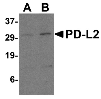
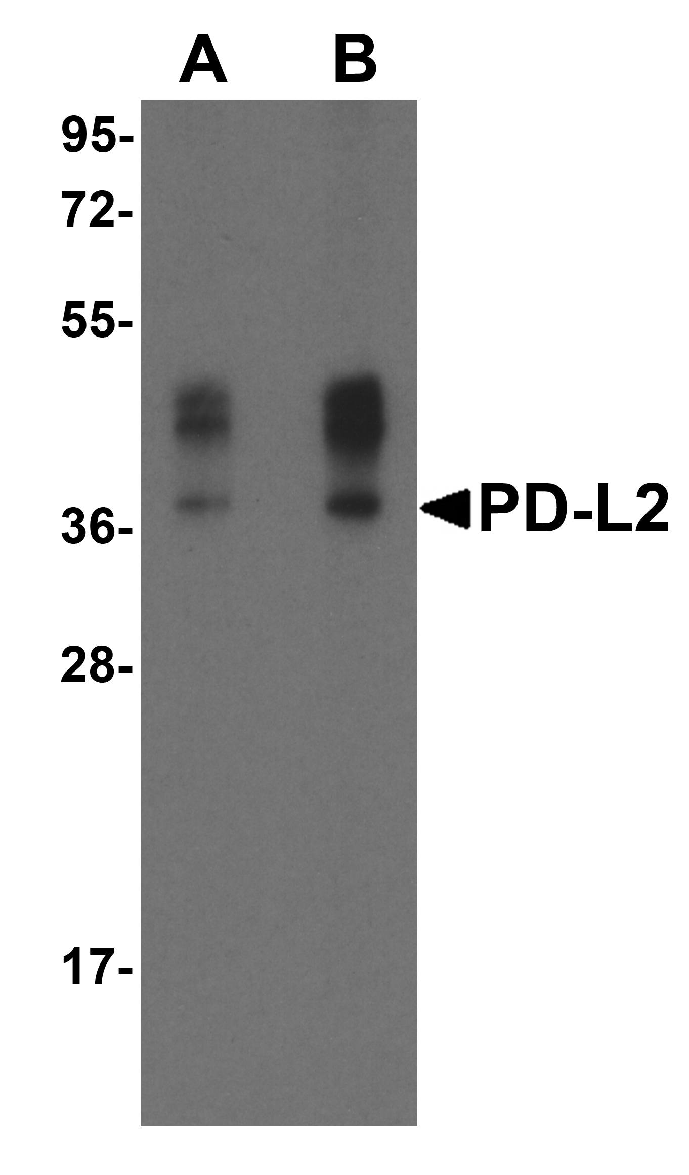
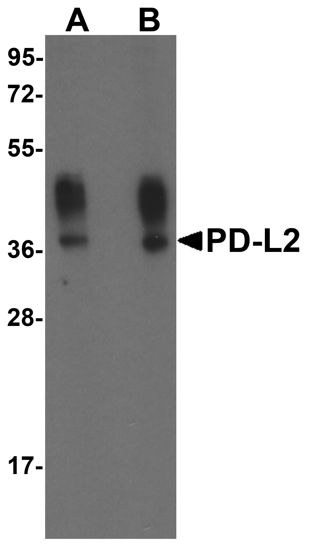
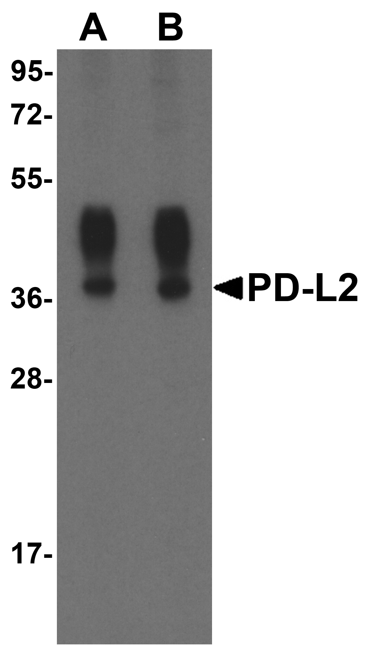
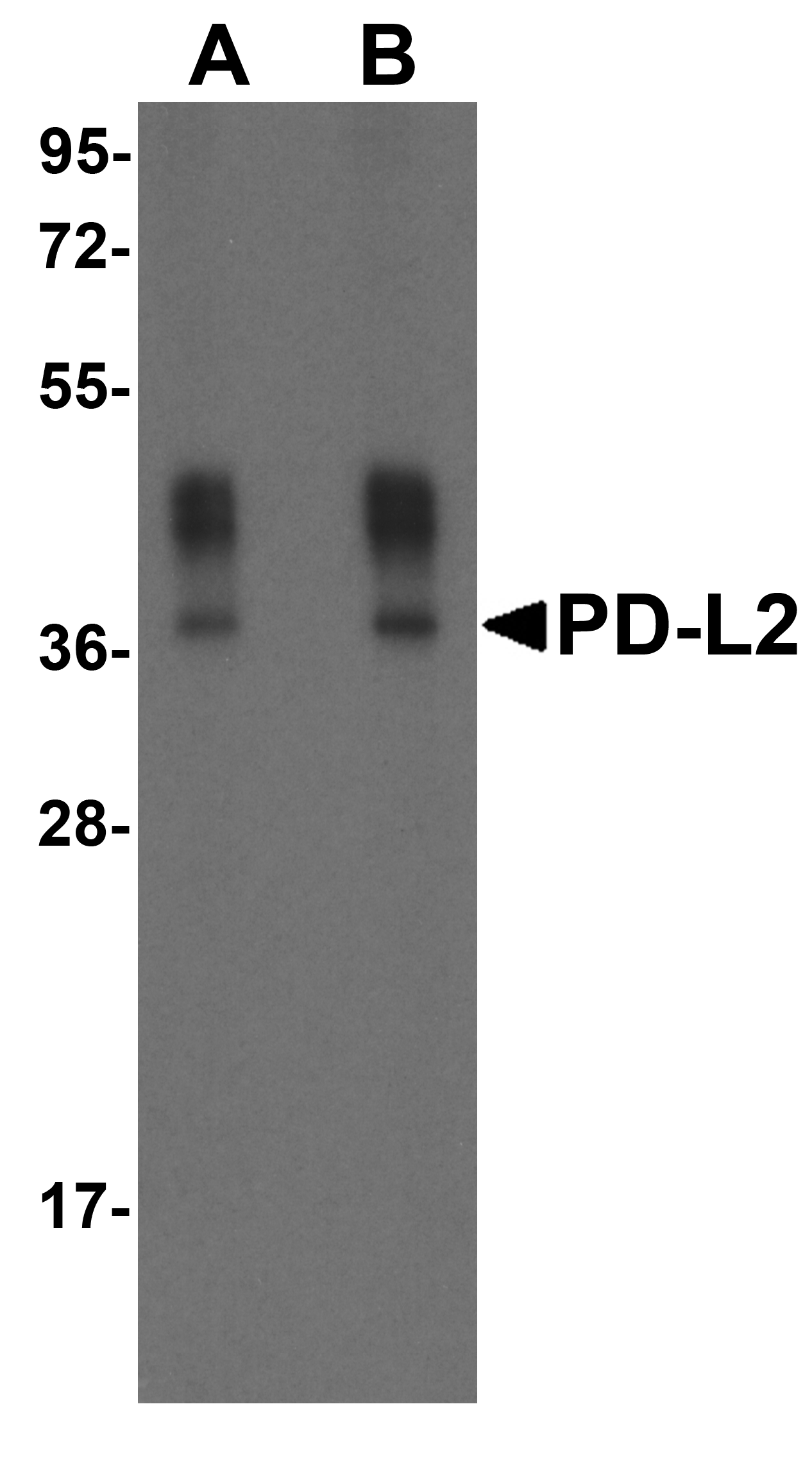
Western Blot Validation in Human Raji Cell Lysate. Loading: 15 µg of lysates per lane. Antibodies: PD-L2 orb1239861 (A: 0.5 µg/mL and B: 1 µg/mL), 1h incubation at RT in 5% NFDM/TBST. Secondary: Goat anti-rabbit IgG HRP conjugate at 1:10000 dilution.
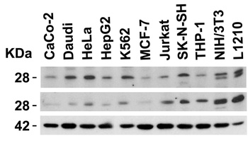
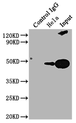
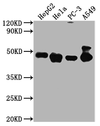
Independent Antibody Validation (IAV) via Protein Expression Profile in Human and Mouse Cell Lines. Loading: 15 µg of lysates per lane. Antibodies: PD-L2, orb1239861 (4 µg/mL), competitor antibody (4 µg/mL), and beta-actin (1 µg/mL), 1h incubation at RT in 5% NFDM/TBST. Secondary: Goat anti-rabbit IgG HRP conjugate at 1:10000 dilution.
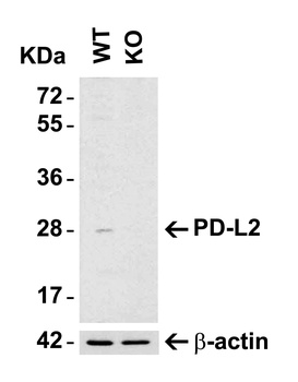
KO Validation in HeLa Cells. Loading: 15 µg of HeLa WT cell lysates or PD-L2 KO cell lysates. Antibodies: PD-L2, orb1239861 (4 µg/mL) and beta-actin orb1240312 (1 µg/mL), 1 h incubation at RT in 5% NFDM/TBST. Secondary: Goat Anti-Rabbit IgG HRP conjugate at 1:10000 dilution.
KO Validation in HeLa Cells. Loading: 15 µg of HeLa WT cell lysates or PD-L2 KO cell lysates. Antibodies: PD-L2, orb1239861 (4 µg/mL) and beta-actin orb1240312 (1 µg/mL), 1 h incubation at RT in 5% NFDM/TBST. Secondary: Goat Anti-Rabbit IgG HRP conjugate at 1:10000 dilution.

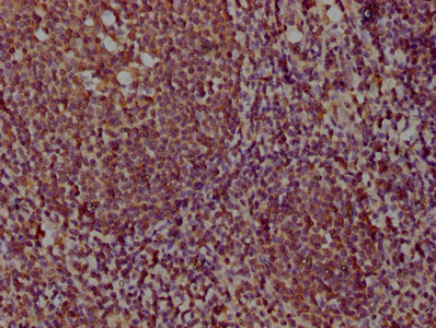
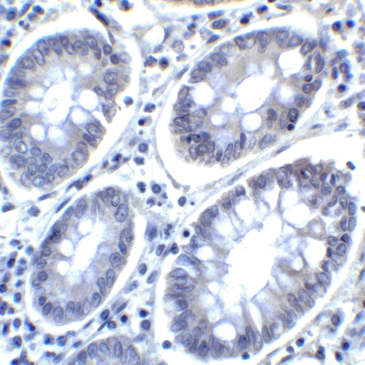
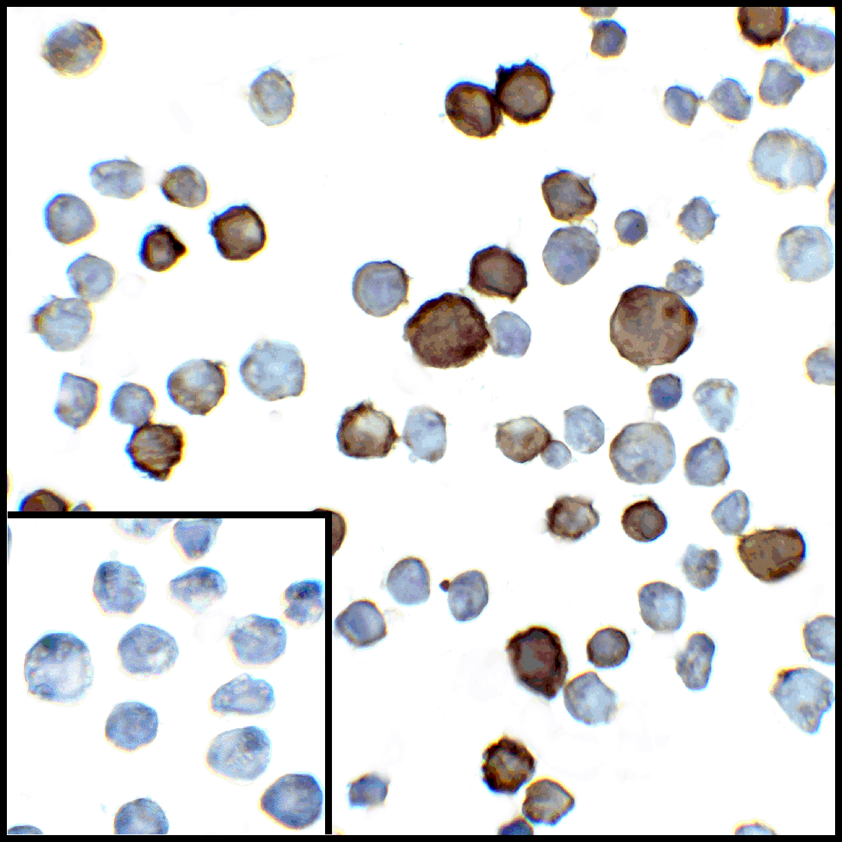
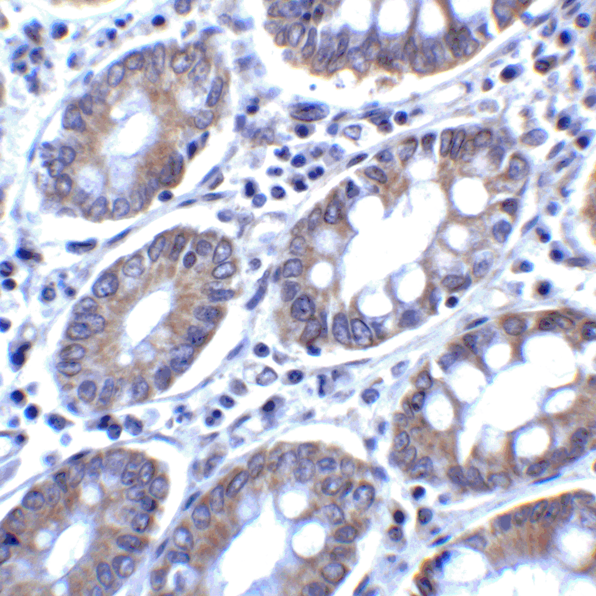
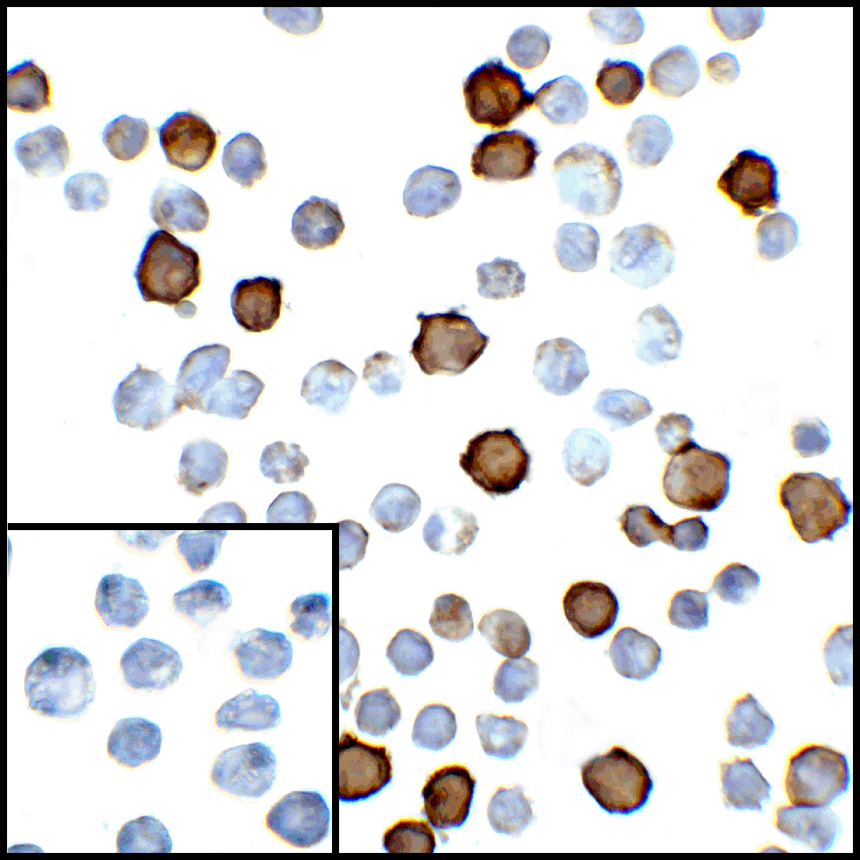
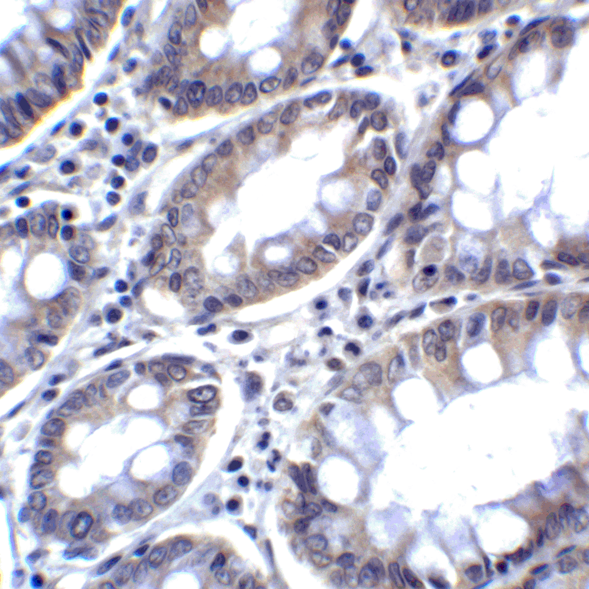
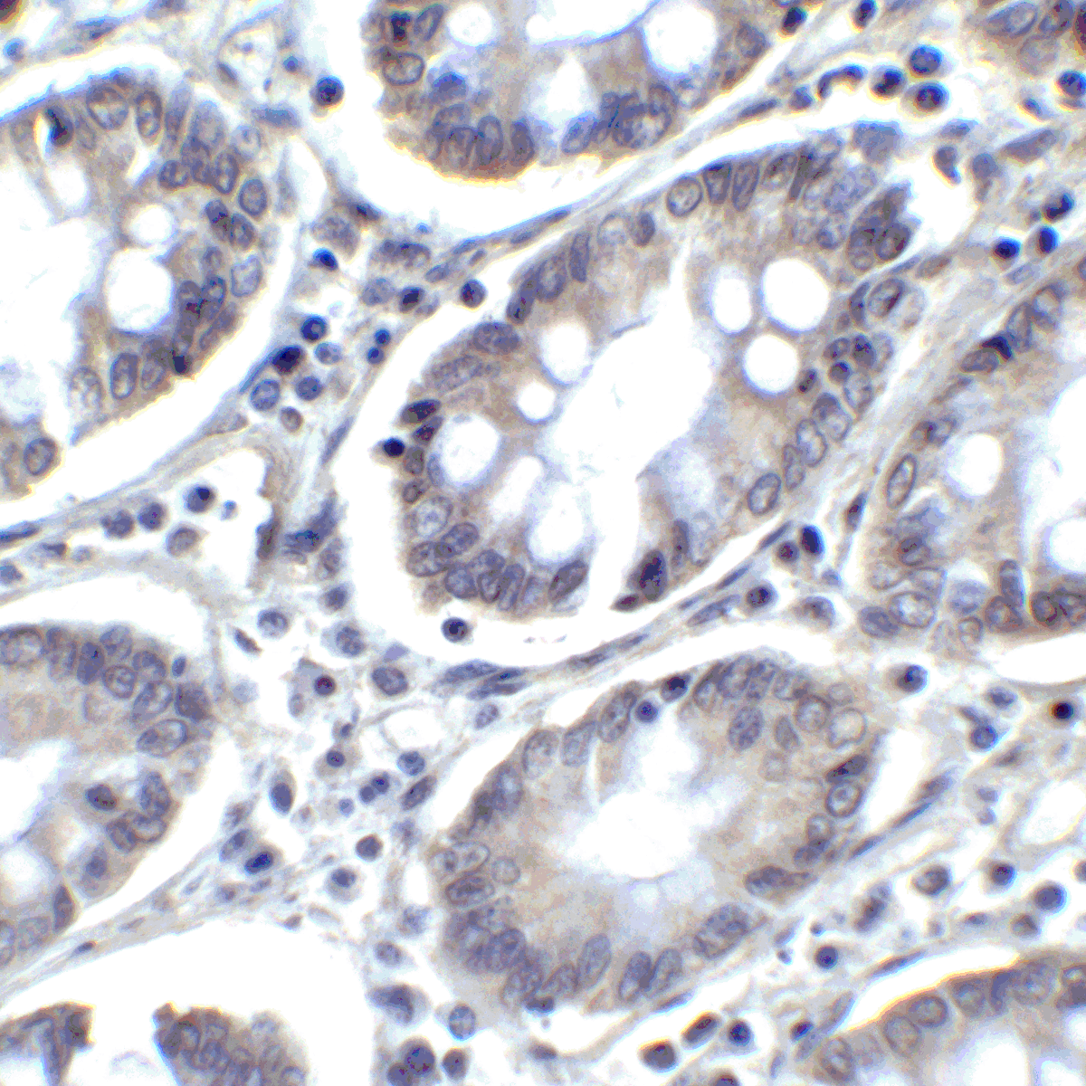
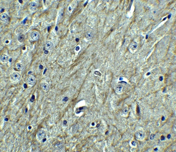
Immunohistochemistry Validation of PD-L2 in Mouse Brain Tissue. Immunohistochemical analysis of paraffin-embedded mouse brain tissue using anti-PD-L2 antibody (orb1239861) at 2.5 µg/ml. Tissue was fixed with formaldehyde and blocked with 10% serum for 1 h at RT; antigen retrieval was by heat mediation with a citrate buffer (pH6). Samples were incubated with primary antibody overnight at 4°C. A goat anti-rabbit IgG H&L (HRP) at 1/250 was used as secondary. Counter stained with Hematoxylin.
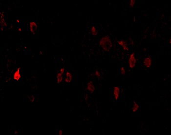
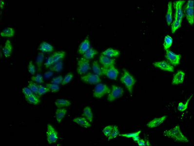
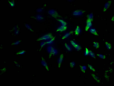
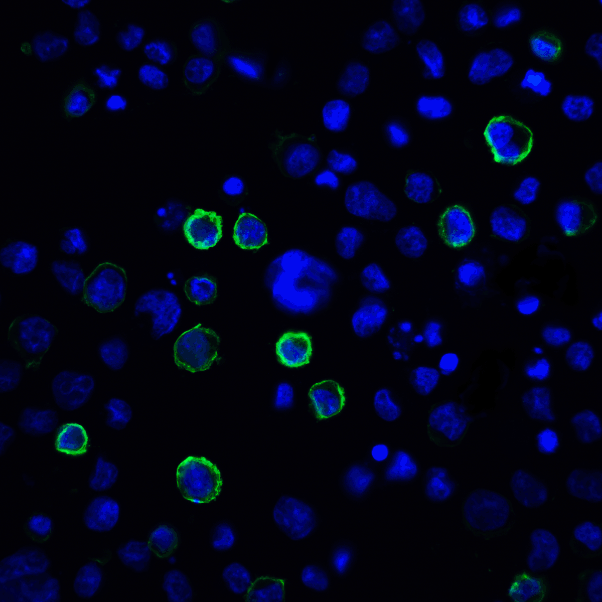
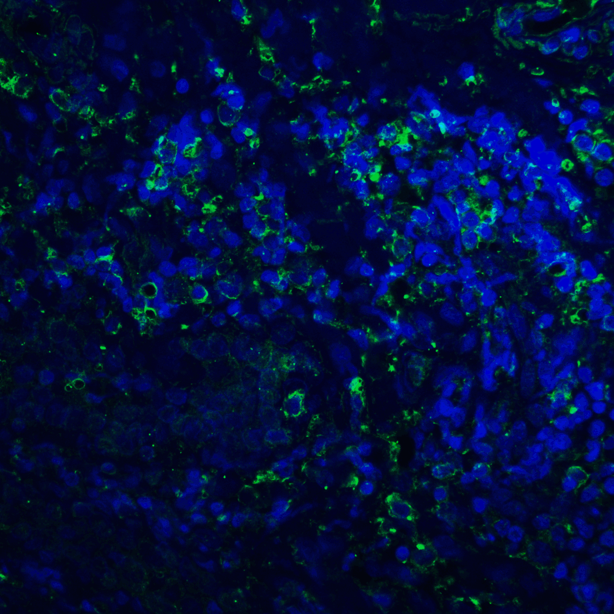
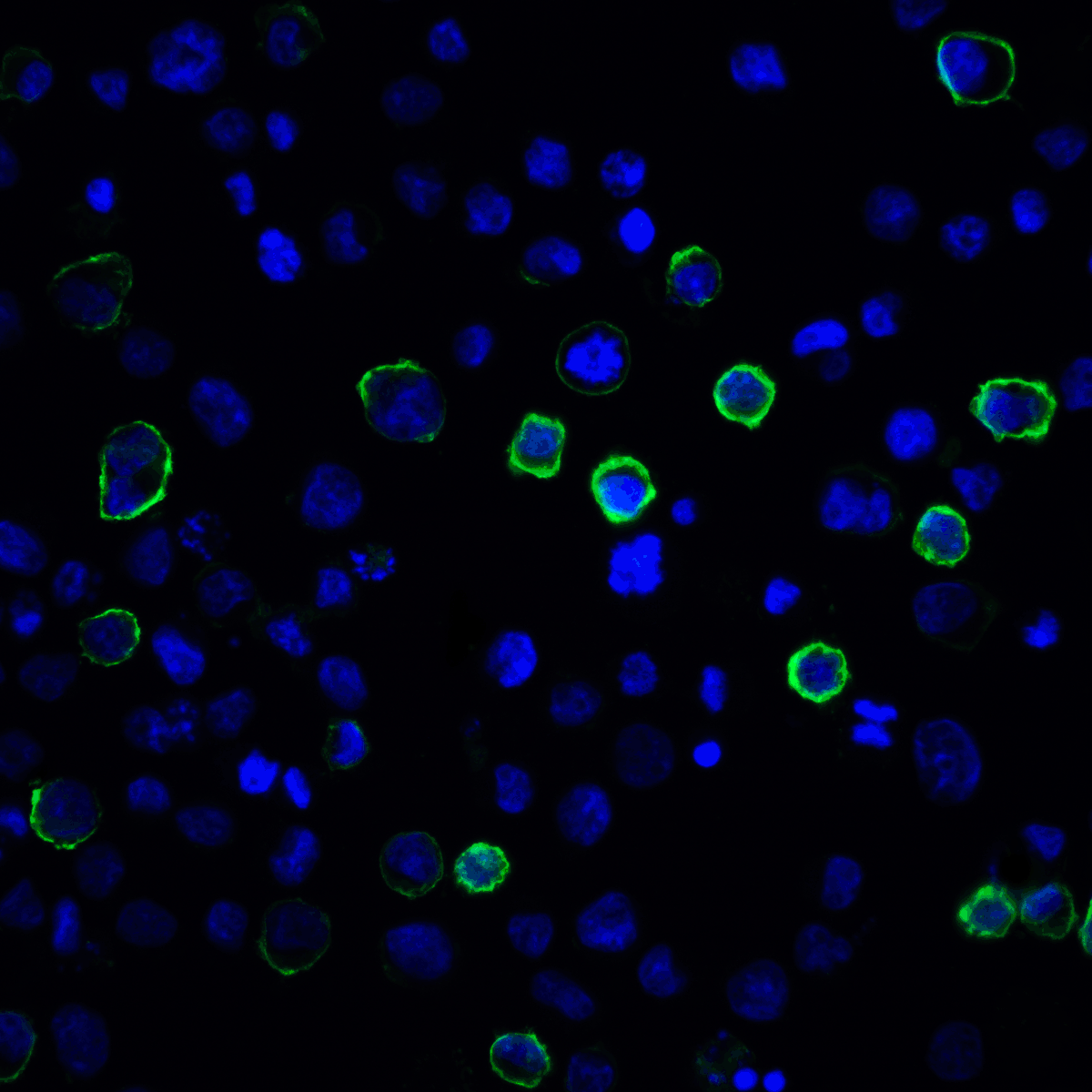
Immunofluorescence Validation of PD-L2 in Mouse Brain Tissue. Immunofluorescent analysis of 4% paraformaldehyde-fixed mouse brain cells labeling PD-L2 with orb1239861 at 20 µg/mL, followed by goat anti-rabbit IgG secondary antibody at 1/500 dilution (red).
Immunohistochemistry Validation of PD-L2 in Mouse Brain Tissue. Immunohistochemical analysis of paraffin-embedded mouse brain tissue using anti-PD-L2 antibody (orb1239861) at 2.5 µg/ml. Tissue was fixed with formaldehyde and blocked with 10% serum for 1 h at RT; antigen retrieval was by heat mediation with a citrate buffer (pH6). Samples were incubated with primary antibody overnight at 4°C. A goat anti-rabbit IgG H&L (HRP) at 1/250 was used as secondary. Counter stained with Hematoxylin.
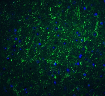
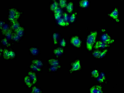
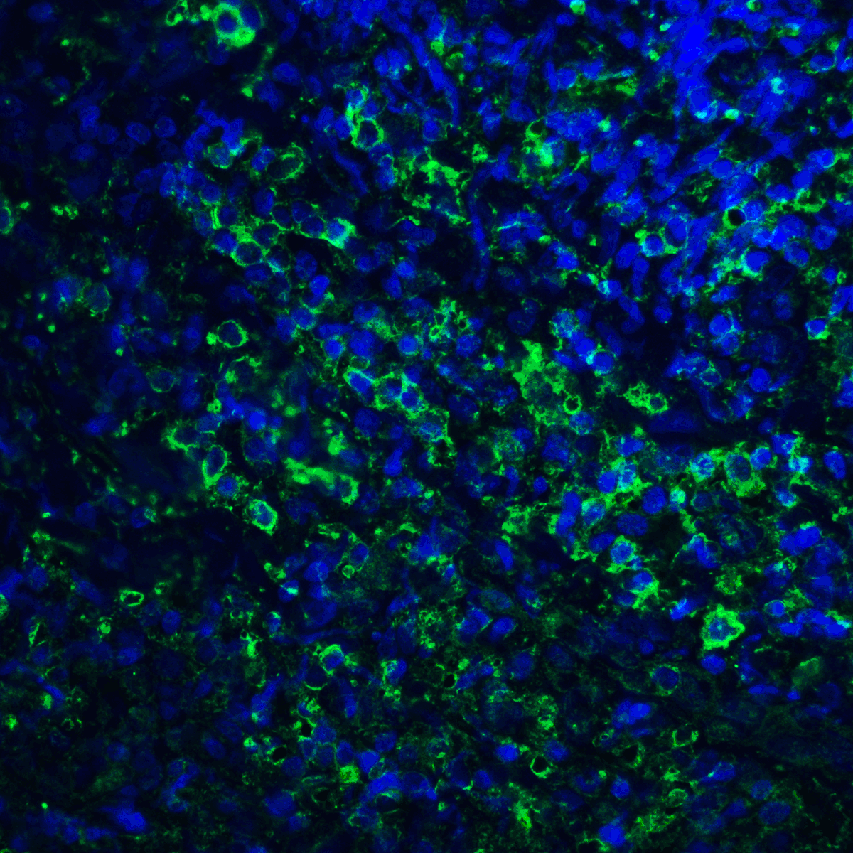
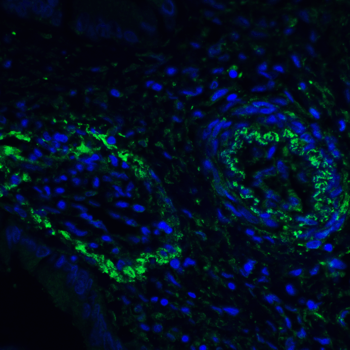
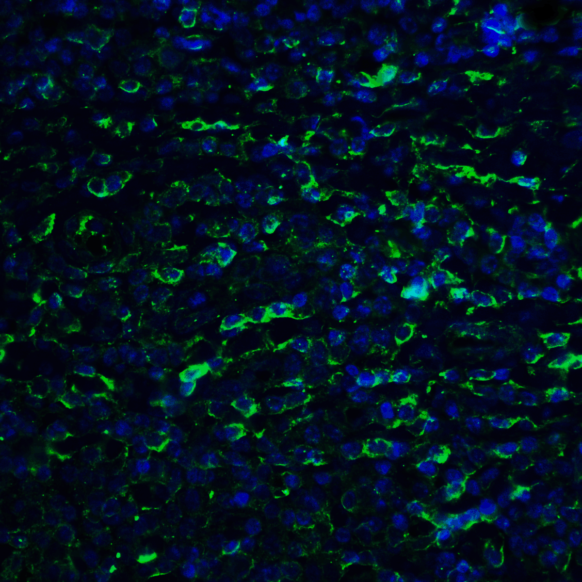
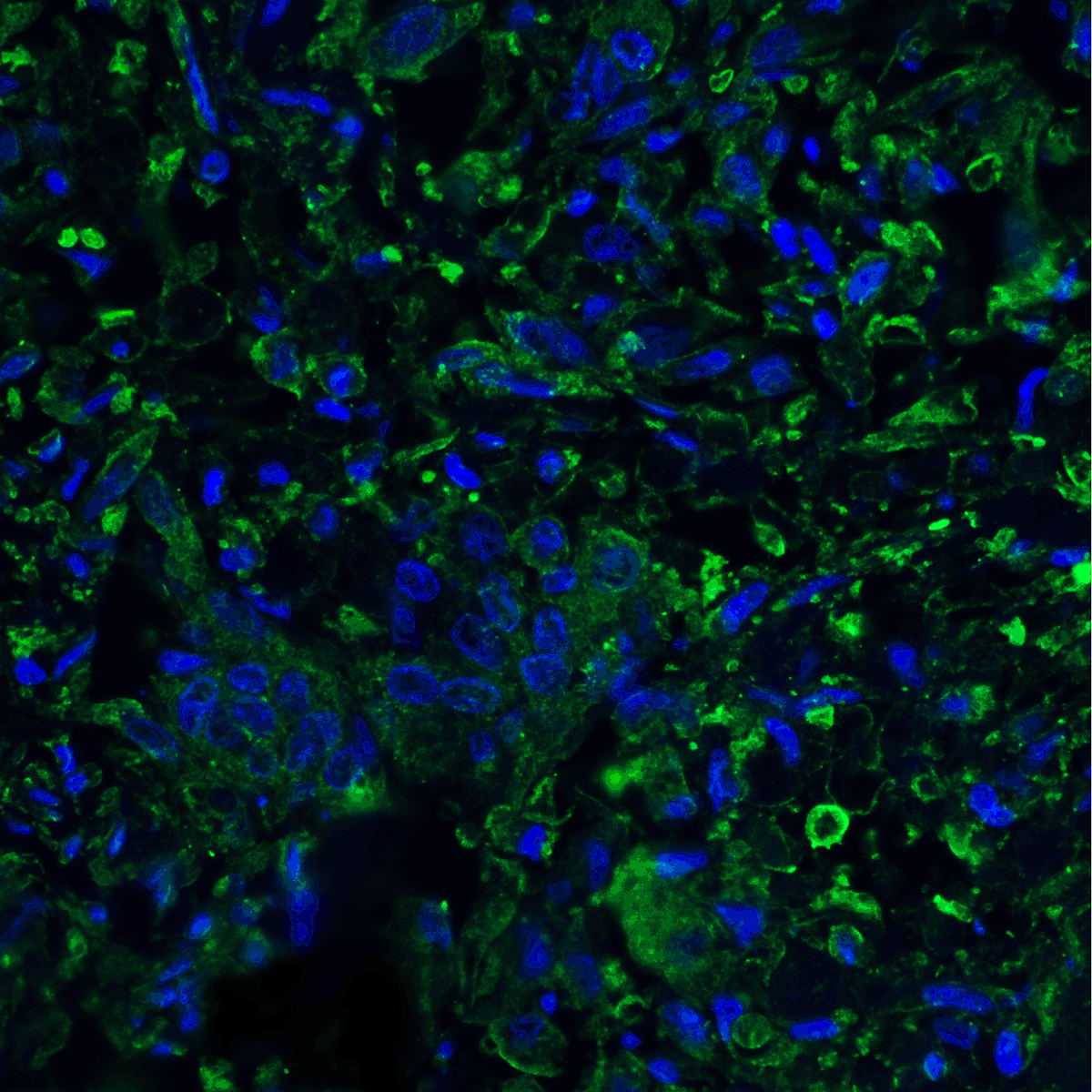
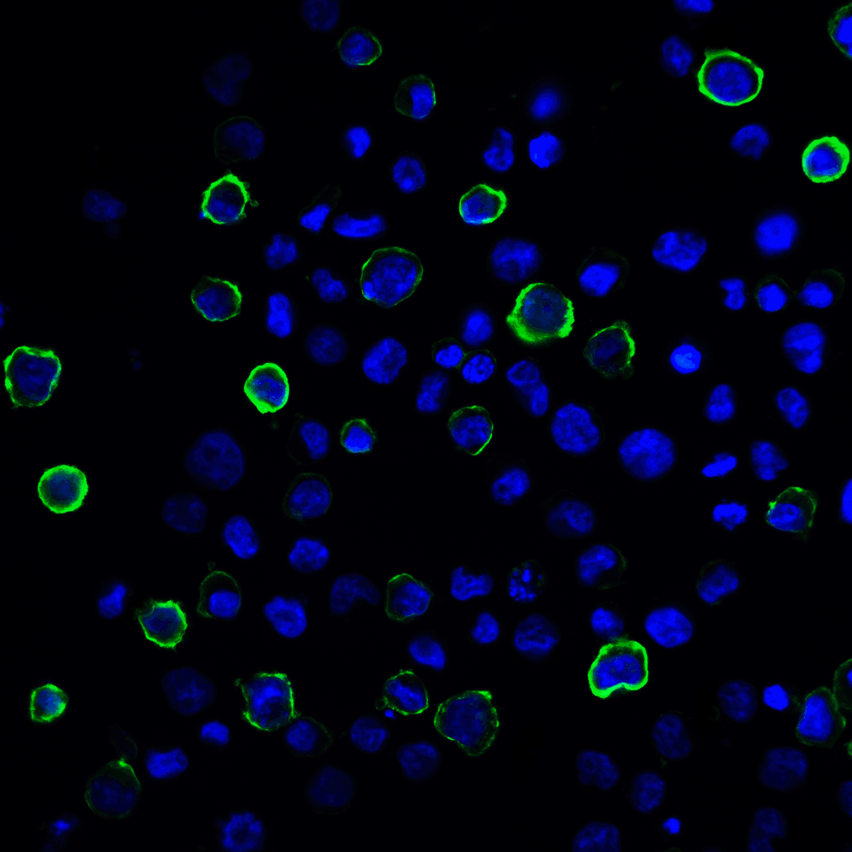
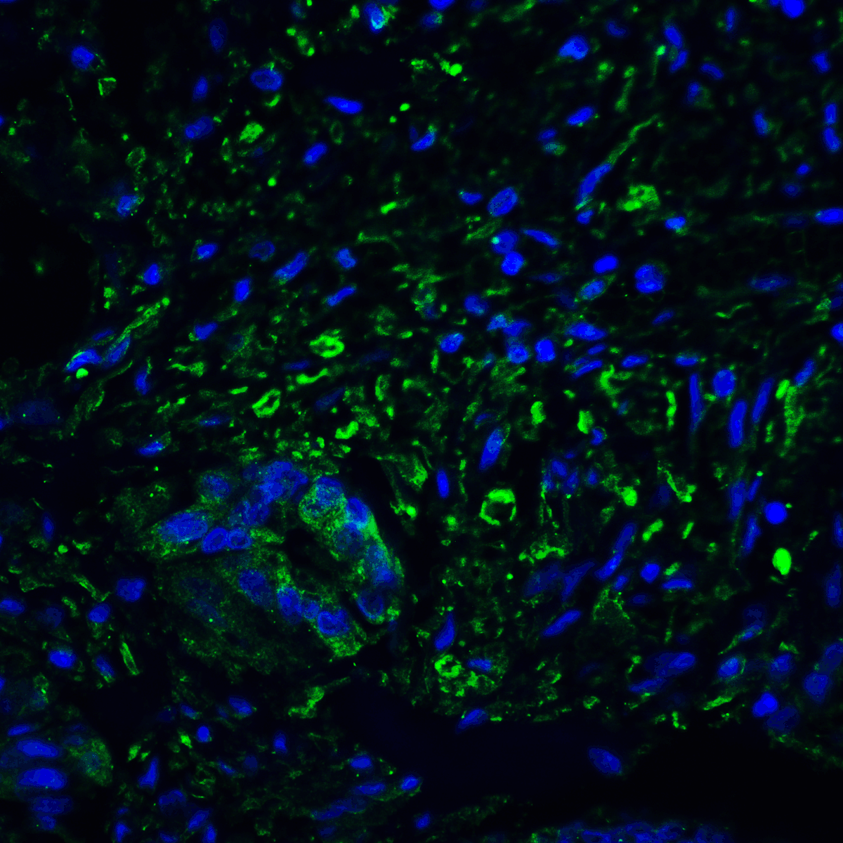
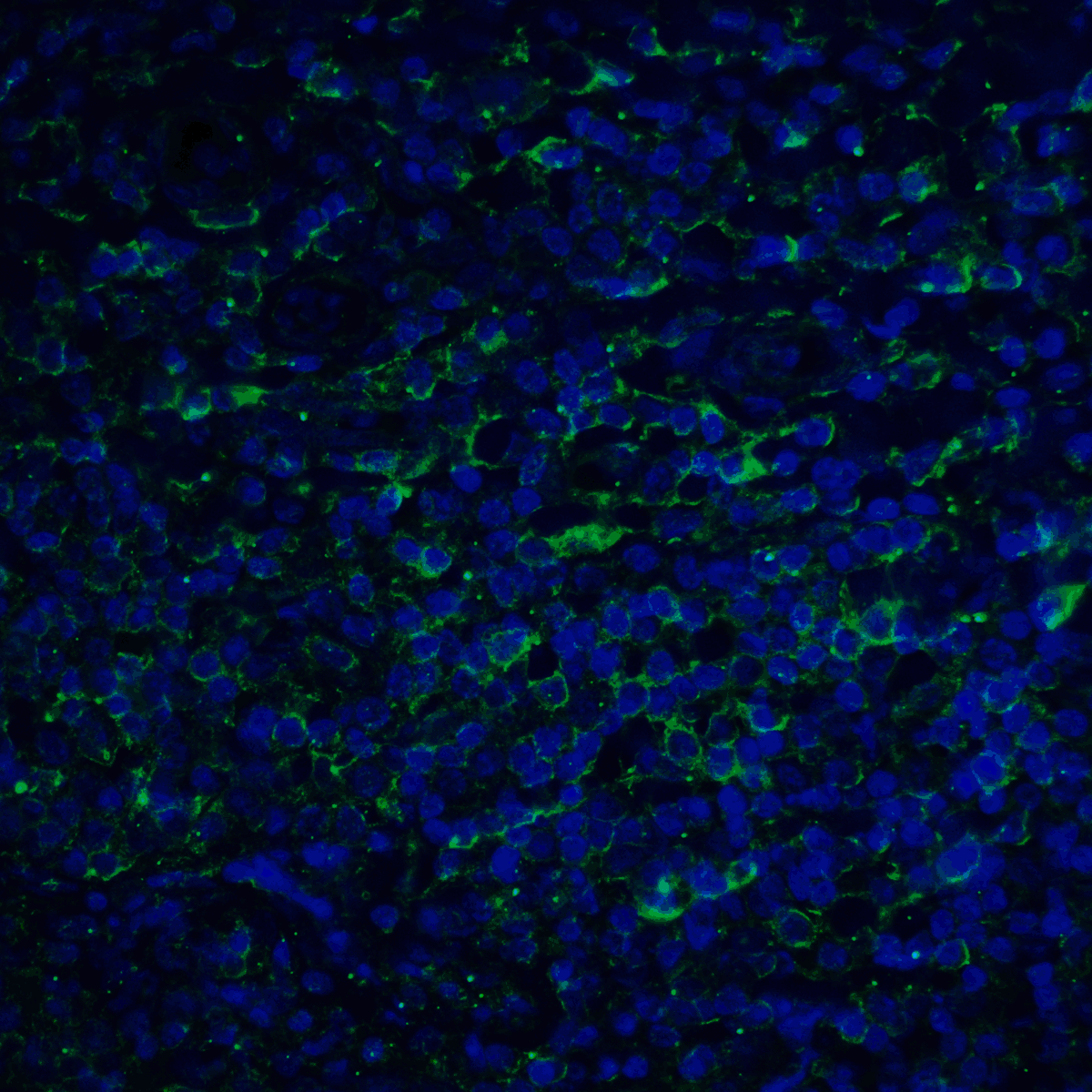
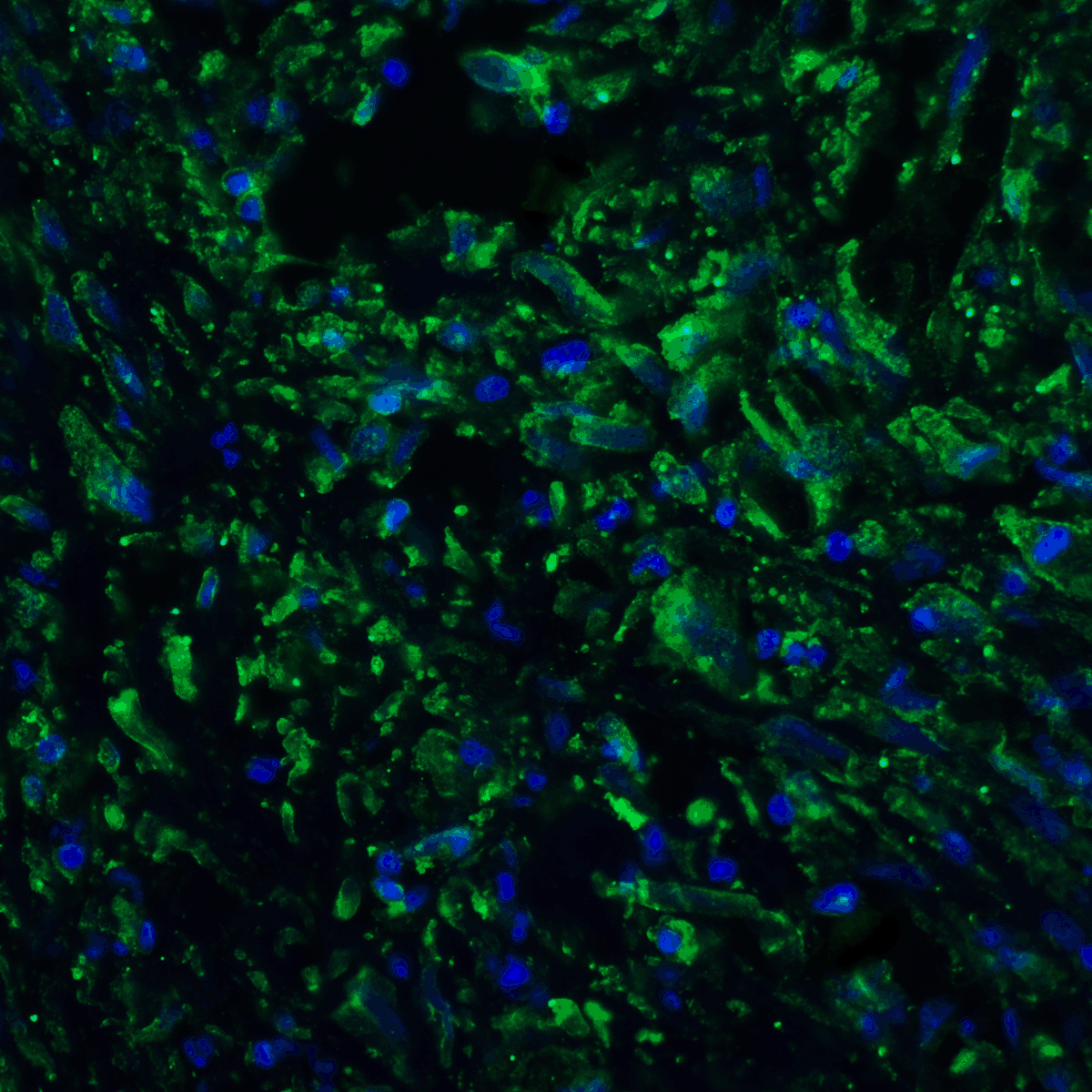
Immunofluorescence Validation of PD-L2 in Mouse Brain Tissue. Immunofluorescent analysis of 4% paraformaldehyde-fixed mouse brain tissue labeling PD-L2 with orb1239861 at 20 µg/mL, followed by goat anti-rabbit IgG secondary antibody at 1/500 dilution (green) and DAPI staining (blue).
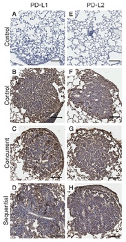
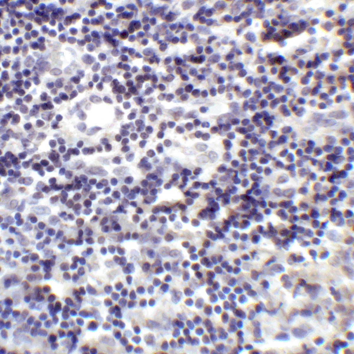
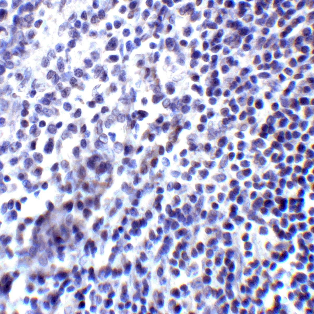
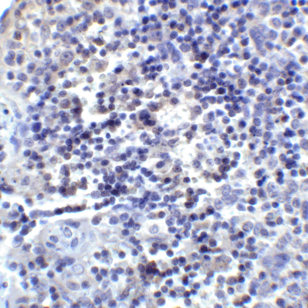
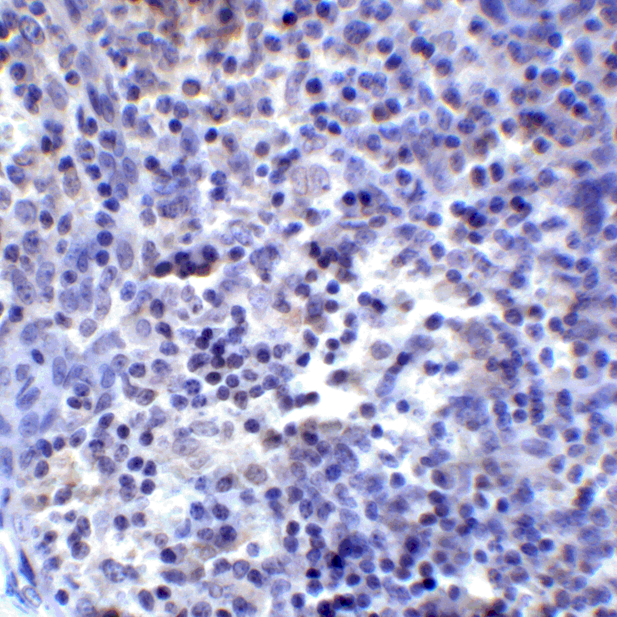
Immunohistochemistry Validation of PD-L2 in Lung Tumor of Mice (Kao et al., 2015). Protein analysis for PD-L2 (E-H) by immunohistochemistry with anti-PD-L2 antibodies in mice lung tumors. hMUC1.Tg mice were induced with lung adenoma and then treated with concurrent or sequential cisplatin/radiotherapy. PD-L2 expression level at week 41 after treatment was similar in control and treatment groups.
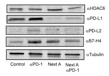
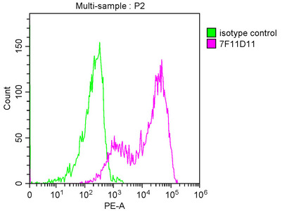
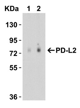
Regulated Expression Validation of PD-L2 in Mice with Melanoma Tumor (Knox et al., 2019). Immunoblot analysis of PD-L2 expression with anti-PD-L2 (orb1239861) antibodies. PD-L2 expression was up-regulated by anti-PD1 antibody treatment whereas it was reduced by Next A alone or combination treatment (anti-PD1 antibody + NextA).
Documents Download
Datasheet
Product Information
Request a Document
Protocol Information
WB
Western Blot (IB, immunoblot)
IHC-P
Immunohistochemistry Paraffin
IF
Immunofluorescence
ELISA
Enzyme-linked Immunosorbent Assay (EIA)
PDCD1LG2 Antibody (orb1239861)

- 0.0
Based on 0 reviews
Participating in our Biorbyt product reviews program enables you to support fellow scientists by sharing your firsthand experience with our products.
Login to Submit a ReviewAvailable Sizes
Select a size below
Free Secondary Antibody (20 ul)0/0
Please add an antibody product to your cart first.