You have no items in your shopping cart.
Description
Research Area
Cancer Biology, Cell Biology, Neuroscience
Images & Validation
−Item 1 of 4
| Tested Applications | IHC-P, WB |
|---|---|
| Dilution Range | WB - 1:1000, IHC-P - 1:50-100 |
| Reactivity | Human, Mouse |
Key Properties
−| Antibody Type | Primary Antibody |
|---|---|
| Host | Rabbit |
| Clonality | Polyclonal |
| Isotype | Rabbit IgG |
| Immunogen | This PBP antibody is generated from rabbits immunized with a KLH conjugated synthetic peptide between 137-167 amino acids from the Central region of human PBP. Antigen Region: 137-167 aa. |
| Target | PEBP1 |
| Molecular Weight | 21057 Da |
| Conjugation | Unconjugated |
Storage & Handling
−| Storage | Maintain refrigerated at 2-8°C for up to 2 weeks. For long term storage store at -20°C in small aliquots to prevent freeze-thaw cycles |
|---|---|
| Form/Appearance | Purified polyclonal antibody supplied in PBS with 0.09% (W/V) sodium azide. This antibody is purified through a protein G column, eluted with high and low pH buffers and neutralized immediately, followed by dialysis against PBS. |
| Expiration Date | 12 months from date of receipt. |
| Disclaimer | For research use only |
Alternative Names
−Phosphatidylethanolamine-binding protein 1, PEBP-1, HCNPpp, Neuropolypeptide h3, Prostatic-binding protein, Raf kinase inhibitor protein, RKIP, Hippocampal cholinergic neurostimulating peptide, HCNP, PEBP1, PBP, PEBP
Similar Products
−- Item 1 of 3
TRAP220 Rabbit Polyclonal Antibody [orb214424]
IF, IHC, WB
Human, Monkey, Mouse, Rat
Rabbit
Polyclonal
Unconjugated
30 μl, 100 μl, 200 μl, 50 μl - Item 1 of 2
RKIP Rabbit Polyclonal Antibody [orb214353]
IH, KO/KD Validated, WB
Human, Mouse, Rat
Rabbit
Polyclonal
Unconjugated
30 μl, 100 μl, 200 μl, 50 μl - Item 1 of 1
PBP Antibody (Center) [orb1787956]
WB
Human
Rabbit
Polyclonal
Unconjugated

Quality Guarantee
Explore bioreagents carefree to elevate your research. All our products are rigorously tested for performance. If a product does not perform as described on its datasheet, our scientific support team will provide expert troubleshooting, a prompt replacement, or a refund. For full details, please see our Terms & Conditions and Buying Guide. Contact us at [email protected].
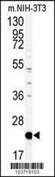
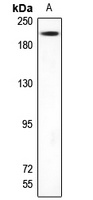
Western blot analysis of anti-PBP Antibody (Center) in mouse NIH-3T3 tissue lysates (35 ug/lane). PBP (arrow) was detected using the purified Pab.
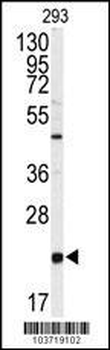
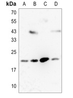
Western blot analysis of anti-PBP Antibody (Center) Antibody (Center) in 293 cell line lysates (35 ug/lane). PBP (arrow) was detected using the purified Pab.
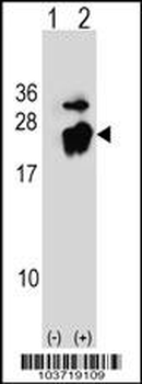
Western blot analysis of PBP (arrow) using rabbit polyclonal PBP Antibody (A152). 293 cell lysates (2 ug/lane) either nontransfected (Lane 1) or transiently transfected (Lane 2) with the PBP gene.
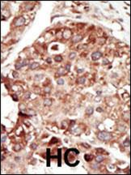
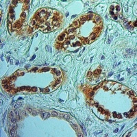
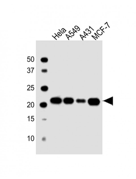
Formalin-fixed and paraffin-embedded human cancer tissue reacted with the primary antibody, which was peroxidase-conjugated to the secondary antibody, followed by DAB staining. This data demonstrates the use of this antibody for immunohistochemistry; clinical relevance has not been evaluated. BC = breast carcinoma; HC = hepatocarcinoma.
Quick Database Links
UniProt Details
− No UniProt data available
NCBI Reference Sequences
−Associated Accession Numbers
Curated reference sequences for the gene transcript and protein product| Protein | NP_002558.1 |
|---|
Documents Download
Datasheet
Product Information
Request a Document
Protocol Information
WB
Western Blot (IB, immunoblot)
IHC-P
Immunohistochemistry Paraffin
PBP Antibody (Center) (orb1928613)

- 0.0
Based on 0 reviews
Participating in our Biorbyt product reviews program enables you to support fellow scientists by sharing your firsthand experience with our products.
Login to Submit a ReviewAvailable Sizes
Select a size below
Choose Conjugation or Carrier Free Version
Free Secondary Antibody (20 ul)0/0
Please add an antibody product to your cart first.