You have no items in your shopping cart.
Description
Research Area
Cardiovascular Research
Images & Validation
−Item 1 of 5
| Tested Applications | DOT, ELISA, IHC, WB |
|---|---|
| Dilution Range | ELISA: 1:20,000 - 1:60,000, IHC: 1:200, WB: 1:500- 1:2,000 |
| Reactivity | Human, Mouse |
| Application Notes |
Key Properties
−| Antibody Type | Primary Antibody |
|---|---|
| Host | Rabbit |
| Clonality | Polyclonal |
| Isotype | Antiserum |
| Immunogen | This whole rabbit serum was prepared by repeated immunizations with a synthetic peptide corresponding to amino acid residues of human Notch 1 located near the N-terminal sequence of the cleaved N intracellular domain (NICD). |
| Target | NOTCH1 |
| Purity | This antiserum is directed against human NOTCH 1. Based on the immunogen sequence, we expect this antibody to react as well with mouse and rat NOTCH 1 (100% sequence homology). This antibody reacts with mouse Notch constructs present in lysates of HEK 293 cells. Only the cleaved intracellular (activated) form (NICD) is detected. No reactivity is detected against mouse N2, N3 or N4. The immunogen epitope is only exposed after gamma secretase cleavage and is not accessible in the uncleaved form. |
| Conjugation | Unconjugated |
Storage & Handling
−| Storage | Store vial at -20° C or below prior to opening. This vial contains a relatively low volume of reagent (25 µL). To minimize loss of volume dilute 1:10 by adding 225 µL of the buffer stated above directly to the vial. Recap, mix thoroughly and briefly centrifuge to collect the volume at the bottom of the vial. Use this intermediate dilution when calculating final dilutions as recommended below. Store the vial at -20°C or below after dilution. Avoid cycles of freezing and thawing. |
|---|---|
| Form/Appearance | Liquid (sterile filtered) |
| Buffer/Preservatives | 0.1% (w/v) Sodium Azide |
| Concentration | 90 mg/mL |
| Expiration Date | 12 months from date of receipt. |
| Dry Ice Shipping | Please note: This product requires shipment on dry ice. A dry ice surcharge will apply. |
| Disclaimer | For research use only |
Alternative Names
−rabbit anti-notch1 antibody, Neurogenic locus Notch homolog protein 1, hN1, Translocation-associated Notch protein TAN-1
Similar Products
−- Item 1 of 17
Notch1 Rabbit Polyclonal Antibody [orb500788]
IF, IHC-Fr, IHC-P, WB
Mouse, Rat
Human, Mouse, Rat
Rabbit
Polyclonal
Unconjugated
50 μl, 100 μl, 200 μl - Item 1 of 7
NOTCH1 Rabbit Polyclonal Antibody [orb577050]
WB
Bovine, Canine, Equine, Guinea pig, Mouse, Rat, Zebrafish
Human
Rabbit
Polyclonal
Unconjugated
100 μl - Item 1 of 4
Activated Notch1 Rabbit Polyclonal Antibody [orb312158]
IF, IHC-Fr, IHC-P, WB
Bovine, Canine, Guinea pig, Human, Porcine, Rabbit, Rat
Mouse
Rabbit
Polyclonal
Unconjugated
50 μl, 100 μl, 200 μl - Item 1 of 4
- Item 1 of 1
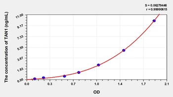
Human Translocation Associated Notch Homolog 1 (TAN1) ELISA Kit [orb779253]
Human
0.16-10 ng/mL
0.055 ng/mL
48 T, 96 T

Quality Guarantee
Explore bioreagents carefree to elevate your research. All our products are rigorously tested for performance. If a product does not perform as described on its datasheet, our scientific support team will provide expert troubleshooting, a prompt replacement, or a refund. For full details, please see our Terms & Conditions and Buying Guide. Contact us at [email protected].
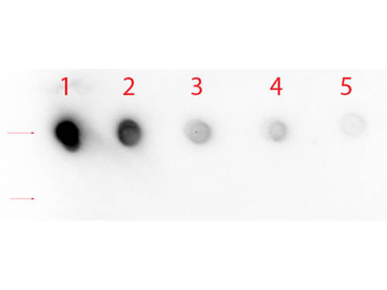
Dot Blot of Rabbit anti-Notch 1 (Cleaved N Terminal) (Human Specific) Antibody. Antigen: Row 1 - Notch 1 Peptide (Cleaved N Terminal) Row 2 - Notch 1 (Intra) Peptide. Load: Lane 1 - 200 ng Lane 2 - 66.67 ng Lane 3 - 22.22 ng Lane 4 - 7.41 ng Lane 5 - 2.47 ng. Primary antibody: Rabbit anti-Notch 1 (Cleaved N Terminal) (Human Specific) Antibody at 1:1000 for 60 min at RT. Secondary antibody: HRP Rabbit Secondary at 1:40000 for 30 min at RT. Block: orb348637 for 1 HR at RT.
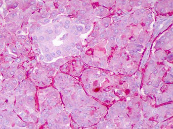
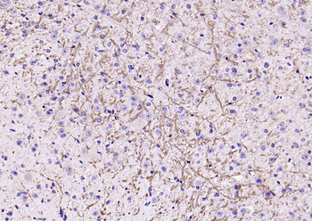
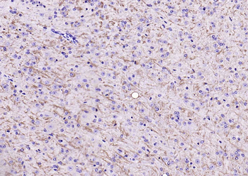
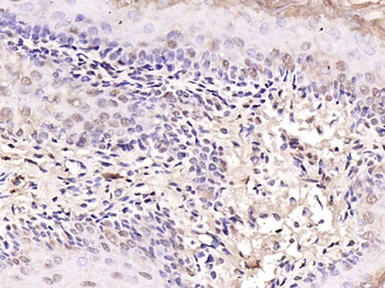
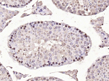
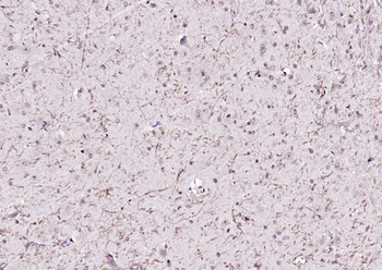
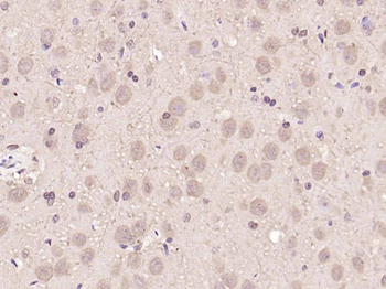
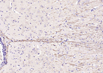
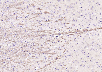
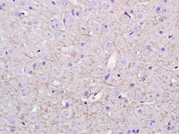
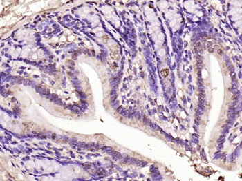
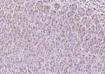
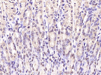
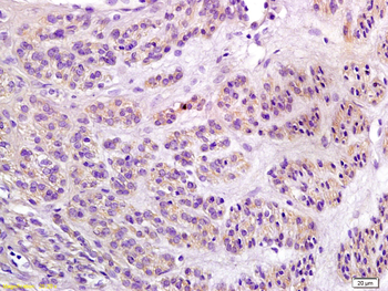
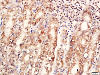
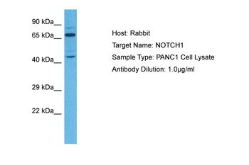
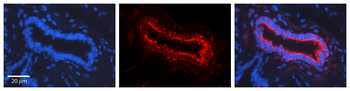
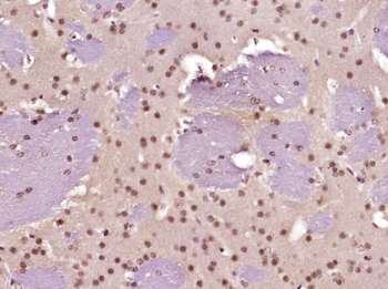
Immunohistochemistry of Rabbit anti-Notch1 antibody. Tissue: Exocrine glands of human pancreas. Fixation: FFPE. Primary antibody: Notch1 antibody at 1:200. Staining: moderate to strong membranous staining and faint to moderate cytoplasmic staining. Islets showed faint staining.
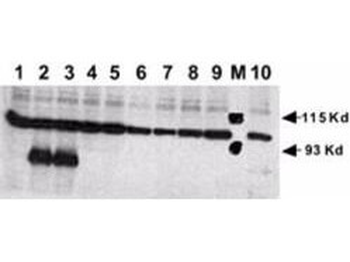
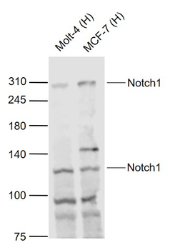
Rabbit anti-Human NOTCH 1 (Cleaved N Terminal) was used at a 1:500 dilution to detect mouse Notch 1 by Western blot. Equivalent amounts of lysates from transiently transfected 293 cells expressing recombinant myc-tagged mouse Notch constructs were electrophoresed and transferred to membrane using standard methods. A reaction with diluted primary antibody was followed by washing; reaction with a 1:10000 dilution of HRP conjugated Gt-a-Rabbit IgG (orb347654), and color development. Lane M: Mol wt markers. Lane 1: No transfection. Lane 2: N1 (mouse deleted extracellular domain)-myc. Lane 3: N1 (mouse intracellular domain)-myc. Lane 4: N2 (mouse deleted extracellular domain)-myc. Lane 5: N2 (mouse intracellular domain)-myc. Lane 6: N3 (mouse deleted extracellular domain)-myc. Lane 7: N3 (mouse intracellular domain)-myc. Lane 8: N4 (mouse deleted extracellular domain)-myc. Lane 9: N4 (mouse intracellular domain)-myc. Lane 10: N1 (mouse deleted extracellular domain)(V to G)-myc.
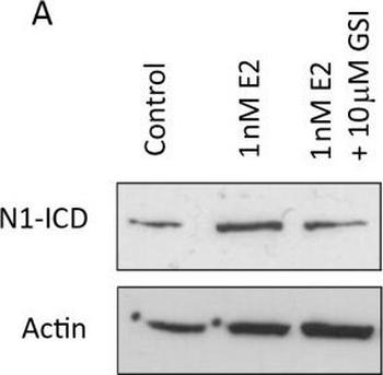
Systemic oestrogen signalling is mediated by EGFR and Notch. (A) Representative Western blot showing expression of cleaved (active) Notch1 (N1-ICD) following culture ± 1 nM 17β-estradiol ± 10 µm GSI. (Bi) Representative Western blot showing expression of Notch ligands in sorted MCF7 cells (left) and, where available, metastatic cells (right). (Bii) Densitometric analysis of three independent repeats of MCF7 sorting and of a single experiment for primary cells. Comparisons between population 1 (CSC enriched) and other populations are displayed. (C and D) Mammosphere formation was assessed following culture with 1 nM 17β-estradiol ± gamma secretase inhibitor (GSI) alone and in combination with gefitinib. Fold change is normalised to control, untreated cells represented as line. (E) Representative image of protein levels of ERK and phosphorylated (actived) ERK following culture for 48 hours in monolayer ± 10 µm GSI. Means plotted ± SEM, *P < 0.05, **P < 0.01, ***P < 0.001 compared to E2 treated. # P < 0.05 compared to control cells.
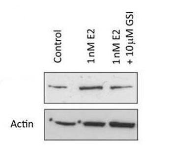
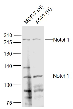
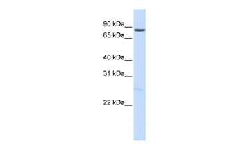
Western Blot of Rabbit anti-Notch1 antibody. Lane 1: MCF-7 control lysate. Lane 2: MCF-7 + 1 nM 17β-estradiol. Lane 3: MCF-7 + 10 µm gamma secretase inhibitor. Load: 35 µg per lane. Primary antibody: Notch1 antibody at 1:500 for overnight at 4°C. Secondary antibody: IRDye800™ rabbit secondary antibody at 1:10000 for 45 min at RT. Block: 5% BLOTTO overnight at 4°C. Predicted/Observed size: 80 kDa for Notch1.
Quick Database Links
UniProt Details
− No UniProt data available
NCBI Reference Sequences
−Associated Accession Numbers
Curated reference sequences for the gene transcript and protein product| Protein | NP_060087.3 |
|---|
Documents Download
Datasheet
Product Information
Request a Document
Protocol Information
WB
Western Blot (IB, immunoblot)
IHC
Immunohistochemistry
ELISA
Enzyme-linked Immunosorbent Assay (EIA)
DOT
Dot Blot
NOTCH1 Antibody (orb750485)

- 0.0
Based on 0 reviews
Participating in our Biorbyt product reviews program enables you to support fellow scientists by sharing your firsthand experience with our products.
Login to Submit a ReviewAvailable Sizes
Select a size below
Free Secondary Antibody (20 ul)0/0
Please add an antibody product to your cart first.